 Movie
Movie Controller

[English] 日本語
 Yorodumi
- PDB-7z03: Endonuclease state of the E. coli Mre11-Rad50 (SbcCD) head comple... -
+ Open data

- Basic information
| Entry | Database: PDB / ID: 7z03 | |||||||||
|---|---|---|---|---|---|---|---|---|---|---|
| Title | Endonuclease state of the E. coli Mre11-Rad50 (SbcCD) head complex bound to ADP and extended dsDNA | |||||||||
 Components Components |
| |||||||||
 Keywords Keywords |  DNA BINDING PROTEIN / ABC-type ATPase / Nuclease / DNA repair DNA BINDING PROTEIN / ABC-type ATPase / Nuclease / DNA repair | |||||||||
| Function / homology |  Function and homology information Function and homology informationdouble-stranded DNA endonuclease activity / DNA exonuclease activity / single-stranded DNA endodeoxyribonuclease activity / 3'-5'-DNA exonuclease activity / DNA repair complex / 3'-5' exonuclease activity / double-strand break repair / DNA recombination / DNA replication / DNA repair ...double-stranded DNA endonuclease activity / DNA exonuclease activity / single-stranded DNA endodeoxyribonuclease activity / 3'-5'-DNA exonuclease activity / DNA repair complex / 3'-5' exonuclease activity / double-strand break repair / DNA recombination / DNA replication / DNA repair / ATP hydrolysis activity / DNA binding / ATP bindingSimilarity search - Function | |||||||||
| Biological species |  Escherichia coli (E. coli) Escherichia coli (E. coli)synthetic construct (others) | |||||||||
| Method | ELECTRON MICROSCOPY / single particle reconstruction / cryo EM / Resolution: 3.7 Å | |||||||||
 Authors Authors | Gut, F. / Kaeshammer, L. / Lammens, K. / Bartho, J. / van de Logt, E. / Kessler, B. / Hopfner, K.P. | |||||||||
| Funding support | European Union,  Germany, 2items Germany, 2items
| |||||||||
 Citation Citation | Journal: Mol Cell / Year: 2022 Title: Structural mechanism of endonucleolytic processing of blocked DNA ends and hairpins by Mre11-Rad50. Authors: Fabian Gut / Lisa Käshammer / Katja Lammens / Joseph D Bartho / Anna-Maria Boggusch / Erik van de Logt / Brigitte Kessler / Karl-Peter Hopfner / Abstract: DNA double-strand breaks (DSBs) threaten genome stability and are linked to tumorigenesis in humans. Repair of DSBs requires the removal of attached proteins and hairpins through a poorly understood ...DNA double-strand breaks (DSBs) threaten genome stability and are linked to tumorigenesis in humans. Repair of DSBs requires the removal of attached proteins and hairpins through a poorly understood but physiologically critical endonuclease activity by the Mre11-Rad50 complex. Here, we report cryoelectron microscopy (cryo-EM) structures of the bacterial Mre11-Rad50 homolog SbcCD bound to a protein-blocked DNA end and a DNA hairpin. The structures reveal that Mre11-Rad50 bends internal DNA for endonucleolytic cleavage and show how internal DNA, DNA ends, and hairpins are processed through a similar ATP-regulated conformational state. Furthermore, Mre11-Rad50 is loaded onto blocked DNA ends with Mre11 pointing away from the block, explaining the distinct biochemistries of 3' → 5' exonucleolytic and endonucleolytic incision through the way Mre11-Rad50 interacts with diverse DNA ends. In summary, our results unify Mre11-Rad50's enigmatic nuclease diversity within a single structural framework and reveal how blocked DNA ends and hairpins are processed. | |||||||||
| History |
|
- Structure visualization
| Structure viewer | Molecule: MolmilJmol/JSmol |
|---|
- Downloads & links
-Download
| PDBx/mmCIF format | 7z03.cif.gz | 366.5 KB | Display | PDBx/mmCIF format |
|---|---|---|---|---|
| PDB format | pdb7z03.ent.gz | 274.2 KB | Display | PDB format |
| PDBx/mmJSON format | 7z03.json.gz | Tree view | PDBx/mmJSON format | |
| Others |  Other downloads Other downloads |
-Validation report
| Arichive directory | https://data.pdbj.org/pub/pdb/validation_reports/z0/7z03ftp://data.pdbj.org/pub/pdb/validation_reports/z0/7z03 | HTTPS FTP |
|---|
-Related structure data
| Related structure data |  14403MC 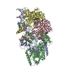 7yzoC  7yzpC C: citing same article ( M: map data used to model this data |
|---|---|
| Similar structure data |


-Links
 PDBj
PDBj


- Assembly
| Deposited unit | 
|
|---|---|
| 1 |
|
-Components
-Nuclease SbcCD subunit ... , 2 types, 4 molecules CDAB
| #1: Protein | Mass: 118851.508 Da / Num. of mol.: 2 Source method: isolated from a genetically manipulated source Source: (gene. exp.) Escherichia coli (E. coli) / Gene: sbcC, rmuA, b0397, JW0387 / Production host: Escherichia coli (E. coli) / References: UniProt: P13458#2: Protein | Mass: 45640.277 Da / Num. of mol.: 2 / Mutation: H84Q Source method: isolated from a genetically manipulated source Source: (gene. exp.) Escherichia coli (E. coli) / Strain: K12 / Gene: sbcD, yajA, b0398, JW0388 / Production host: Escherichia coli (E. coli) / References: UniProt: P0AG76 |
|---|
-DNA chain , 2 types, 2 molecules EF
| #3: DNA chain | Mass: 37099.617 Da / Num. of mol.: 1 / Source method: obtained synthetically / Source: (synth.) synthetic construct (others) / References: GenBank: 1825215262 |
|---|---|
| #4: DNA chain | Mass: 36970.527 Da / Num. of mol.: 1 / Source method: obtained synthetically / Source: (synth.) synthetic construct (others) / References: GenBank: 1825215262 |
-Non-polymers , 3 types, 8 molecules 


| #5: Chemical | Adenosine diphosphate Mass: 427.201 Da / Num. of mol.: 2 / Source method: obtained synthetically / Formula: C10H15N5O10P2 / Feature type: SUBJECT OF INVESTIGATION / Comment: ADP, energy-carrying molecule*YM Mass: 427.201 Da / Num. of mol.: 2 / Source method: obtained synthetically / Formula: C10H15N5O10P2 / Feature type: SUBJECT OF INVESTIGATION / Comment: ADP, energy-carrying molecule*YM#6: Chemical |  Mass: 24.305 Da / Num. of mol.: 2 / Source method: obtained synthetically / Formula: Mg Mass: 24.305 Da / Num. of mol.: 2 / Source method: obtained synthetically / Formula: Mg#7: Chemical | ChemComp-MN /  Mass: 54.938 Da / Num. of mol.: 4 / Source method: obtained synthetically / Formula: Mn Mass: 54.938 Da / Num. of mol.: 4 / Source method: obtained synthetically / Formula: Mn |
|---|
-Details
| Has ligand of interest | Y |
|---|
-Experimental details
-Experiment
| Experiment | Method: ELECTRON MICROSCOPY |
|---|---|
| EM experiment | Aggregation state: PARTICLE / 3D reconstruction method: single particle reconstruction |
- Sample preparation
| Component | Name: Complex of Mre11 dimer, Rad50 dimer and double-stranded DNA Type: COMPLEX / Entity ID: #1-#4 / Source: RECOMBINANT |
|---|---|
| Source (natural) | Organism: Escherichia coli (E. coli) |
| Source (recombinant) | Organism: Escherichia coli (E. coli) |
| Buffer solution | pH: 7.5 |
| Specimen | Embedding applied: NO / Shadowing applied: NO / Staining applied: NO / Vitrification applied: YES |
| Vitrification | Cryogen name: ETHANE |
- Electron microscopy imaging
| Experimental equipment |  Model: Titan Krios / Image courtesy: FEI Company |
|---|---|
| Microscopy | Model: FEI TITAN KRIOS |
| Electron gun | Electron source: FIELD EMISSION GUN / Accelerating voltage: 300 kV / Illumination mode: FLOOD BEAM |
| Electron lens | Mode: BRIGHT FIELDBright-field microscopy / Nominal defocus max: 2800 nm / Nominal defocus min: 1200 nm |
| Image recording | Electron dose: 43.19 e/Å2 / Detector mode: COUNTING / Film or detector model: GATAN K2 SUMMIT (4k x 4k) |
- Processing
| CTF correction | Type: PHASE FLIPPING AND AMPLITUDE CORRECTION |
|---|---|
| 3D reconstruction | Resolution: 3.7 Å / Resolution method: FSC 0.143 CUT-OFF / Num. of particles: 57747 / Symmetry type: POINT |
