Movie
Movie Controller


+ Open data

- Basic information
| Entry | Database: PDB / ID: 7yk5 | |||||||||
|---|---|---|---|---|---|---|---|---|---|---|
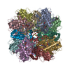
| Title | Rubisco from Phaeodactylum tricornutum bound to PYCO1(452-592) | |||||||||
 Components Components |
| |||||||||
 Keywords Keywords |  PHOTOSYNTHESIS / Rubisco / phase separation / rubisco linker protein / condensation / pyrenoid / phaeodactylum tricornutum PHOTOSYNTHESIS / Rubisco / phase separation / rubisco linker protein / condensation / pyrenoid / phaeodactylum tricornutum | |||||||||
| Function / homology |  Function and homology informationribulose-bisphosphate carboxylase / ribulose-bisphosphate carboxylase activity / reductive pentose-phosphate cycle / chloroplast / monooxygenase activity / magnesium ion binding Function and homology informationribulose-bisphosphate carboxylase / ribulose-bisphosphate carboxylase activity / reductive pentose-phosphate cycle / chloroplast / monooxygenase activity / magnesium ion bindingSimilarity search - Function | |||||||||
| Biological species |  Phaeodactylum tricornutum (Diatom) Phaeodactylum tricornutum (Diatom) | |||||||||
| Method | ELECTRON MICROSCOPY / single particle reconstruction / cryo EM / Resolution: 2 Å | |||||||||
 Authors Authors | Oh, Z.G. / Ang, W.S.L. / Bhushan, S. / Mueller-Cajar, O. | |||||||||
| Funding support |  Singapore, 2items Singapore, 2items
| |||||||||
 Citation Citation | Journal: Proc Natl Acad Sci U S A / Year: 2023 Title: A linker protein from a red-type pyrenoid phase separates with Rubisco via oligomerizing sticker motifs. Authors: Zhen Guo Oh / Warren Shou Leong Ang / Cheng Wei Poh / Soak-Kuan Lai / Siu Kwan Sze / Hoi-Yeung Li / Shashi Bhushan / Tobias Wunder / Oliver Mueller-Cajar / Abstract: The slow kinetics and poor substrate specificity of the key photosynthetic CO-fixing enzyme Rubisco have prompted the repeated evolution of Rubisco-containing biomolecular condensates known as ...The slow kinetics and poor substrate specificity of the key photosynthetic CO-fixing enzyme Rubisco have prompted the repeated evolution of Rubisco-containing biomolecular condensates known as pyrenoids in the majority of eukaryotic microalgae. Diatoms dominate marine photosynthesis, but the interactions underlying their pyrenoids are unknown. Here, we identify and characterize the Rubisco linker protein PYCO1 from . PYCO1 is a tandem repeat protein containing prion-like domains that localizes to the pyrenoid. It undergoes homotypic liquid-liquid phase separation (LLPS) to form condensates that specifically partition diatom Rubisco. Saturation of PYCO1 condensates with Rubisco greatly reduces the mobility of droplet components. Cryo-electron microscopy and mutagenesis data revealed the sticker motifs required for homotypic and heterotypic phase separation. Our data indicate that the PYCO1-Rubisco network is cross-linked by PYCO1 stickers that oligomerize to bind to the small subunits lining the central solvent channel of the Rubisco holoenzyme. A second sticker motif binds to the large subunit. Pyrenoidal Rubisco condensates are highly diverse and tractable models of functional LLPS. | |||||||||
| History |
|
- Structure visualization
| Structure viewer | Molecule: MolmilJmol/JSmol |
|---|
- Downloads & links
-Download
| PDBx/mmCIF format | 7yk5.cif.gz | 838.7 KB | Display | PDBx/mmCIF format |
|---|---|---|---|---|
| PDB format | pdb7yk5.ent.gz | 714.2 KB | Display | PDB format |
| PDBx/mmJSON format | 7yk5.json.gz | Tree view | PDBx/mmJSON format | |
| Others |  Other downloads Other downloads |
-Validation report
| Arichive directory | https://data.pdbj.org/pub/pdb/validation_reports/yk/7yk5ftp://data.pdbj.org/pub/pdb/validation_reports/yk/7yk5 | HTTPS FTP |
|---|
-Related structure data
| Related structure data |  33887MC M: map data used to model this data C: citing same article ( |
|---|---|
| Similar structure data |


-Links
 PDBj
PDBj
- Assembly
| Deposited unit | 
|
|---|---|
| 1 |
|
-Components
| #1: Protein | Mass: 54244.527 Da / Num. of mol.: 8 / Source method: isolated from a natural source / Source: (natural) Phaeodactylum tricornutum (Diatom) / Plasmid details: Pt 1 8.6 CCMP 2561References: UniProt: E9PAI6, ribulose-bisphosphate carboxylase#2: Protein | Mass: 16039.053 Da / Num. of mol.: 8 / Source method: isolated from a natural source / Source: (natural) Phaeodactylum tricornutum (Diatom) / Plasmid details: Pt 1 8.6 CCMP 2561 / References: UniProt: A0A6B9XNC0#3: Protein/peptide | Mass: 875.973 Da / Num. of mol.: 4 Source method: isolated from a genetically manipulated source Details: Pt 1 8.6 CCMP 2561 / Source: (gene. exp.) Phaeodactylum tricornutum (Diatom)Production host:  Escherichia coli 'BL21-Gold(DE3)pLysS AG' (bacteria) Escherichia coli 'BL21-Gold(DE3)pLysS AG' (bacteria)#4: Protein/peptide | Mass: 993.051 Da / Num. of mol.: 8 Source method: isolated from a genetically manipulated source Details: Pt 1 8.6 CCMP 2561 / Source: (gene. exp.) Phaeodactylum tricornutum (Diatom)Production host: Escherichia coli 'BL21-Gold(DE3)pLysS AG' (bacteria)#5: Sugar | ChemComp-CAP /   Type: saccharideCarbohydrate / Mass: 356.115 Da / Num. of mol.: 8 / Source method: obtained synthetically / Formula: C6H14O13P2 / Feature type: SUBJECT OF INVESTIGATION Type: saccharideCarbohydrate / Mass: 356.115 Da / Num. of mol.: 8 / Source method: obtained synthetically / Formula: C6H14O13P2 / Feature type: SUBJECT OF INVESTIGATIONHas ligand of interest | Y | |
|---|
-Experimental details
-Experiment
| Experiment | Method: ELECTRON MICROSCOPY |
|---|---|
| EM experiment | Aggregation state: PARTICLE / 3D reconstruction method: single particle reconstruction |
- Sample preparation
| Component | Name: Rubisco from Phaeodactylum tricornutum bound to linker protein PYCO1 Type: COMPLEX Details: Rubisco purified from source organism, PYCO1 purified from E. coli. Entity ID: #1-#4 / Source: MULTIPLE SOURCES | |||||||||||||||
|---|---|---|---|---|---|---|---|---|---|---|---|---|---|---|---|---|
| Molecular weight | Value: 550 kDa/nm / Experimental value: YES | |||||||||||||||
| Source (natural) | Organism: Phaeodactylum tricornutum (Diatom) | |||||||||||||||
| Buffer solution | pH: 8 / Details: 20 mM Tris pH 8.0 20 mM NaCl | |||||||||||||||
| Buffer component |
| |||||||||||||||
| Specimen | Conc.: 0.5 mg/ml / Embedding applied: NO / Shadowing applied: NO / Staining applied: NO / Vitrification applied: YES Details: 0.5 mg/mL Rubisco incubated with 21.9 uM of PYCO1(452-592). | |||||||||||||||
| Specimen support | Grid material: COPPER / Grid mesh size: 200 divisions/in. / Grid type: Quantifoil R2/2 | |||||||||||||||
| Vitrification | Instrument: FEI VITROBOT MARK IV / Cryogen name: ETHANE / Humidity: 100 % / Chamber temperature: 277 K / Details: Blotted for 2 sec with blot force of 1. |
- Electron microscopy imaging
| Experimental equipment |  Model: Titan Krios / Image courtesy: FEI Company |
|---|---|
| Microscopy | Model: FEI TITAN KRIOS |
| Electron gun | Electron source: FIELD EMISSION GUN / Accelerating voltage: 300 kV / Illumination mode: FLOOD BEAM |
| Electron lens | Mode: BRIGHT FIELDBright-field microscopy / Nominal magnification: 165000 X / Nominal defocus max: 1600 nm / Nominal defocus min: 800 nm / Cs: 2.7 mm / C2 aperture diameter: 100 µm / Alignment procedure: COMA FREE |
| Specimen holder | Cryogen: NITROGEN / Specimen holder model: FEI TITAN KRIOS AUTOGRID HOLDER |
| Image recording | Average exposure time: 5 sec. / Electron dose: 65 e/Å2 / Film or detector model: GATAN K3 (6k x 4k) / Num. of grids imaged: 1 / Num. of real images: 8861 Details: Images were collected in movie mode at 10 frames per second. |
| EM imaging optics | Energyfilter name: GIF Bioquantum / Details: Gatan EF / Energyfilter slit width: 20 eV |
| Image scans | Width: 5760 / Height: 4092 |
- Processing
| EM software |
| ||||||||||||||||||||||||||||||||||||||||||||||||||
|---|---|---|---|---|---|---|---|---|---|---|---|---|---|---|---|---|---|---|---|---|---|---|---|---|---|---|---|---|---|---|---|---|---|---|---|---|---|---|---|---|---|---|---|---|---|---|---|---|---|---|---|
| CTF correction | Type: PHASE FLIPPING ONLY | ||||||||||||||||||||||||||||||||||||||||||||||||||
| Particle selection | Num. of particles selected: 3354751 | ||||||||||||||||||||||||||||||||||||||||||||||||||
| Symmetry | Point symmetry: D4 (2x4 fold dihedral) | ||||||||||||||||||||||||||||||||||||||||||||||||||
| 3D reconstruction | Resolution: 2 Å / Resolution method: FSC 0.143 CUT-OFF / Num. of particles: 259796 / Algorithm: BACK PROJECTION / Num. of class averages: 1 / Symmetry type: POINT | ||||||||||||||||||||||||||||||||||||||||||||||||||
| Atomic model building | Protocol: RIGID BODY FIT / Space: REAL / Details: Coot was used for model building | ||||||||||||||||||||||||||||||||||||||||||||||||||
| Atomic model building | PDB-ID: 5MZ2 |