 Movie
Movie Controller

[English] 日本語
 Yorodumi
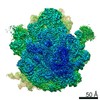
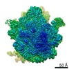
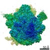
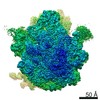
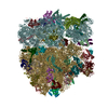
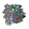
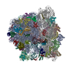
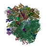
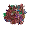
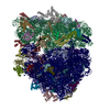
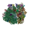
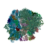
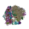
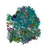
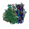
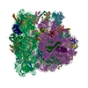
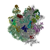
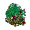
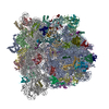
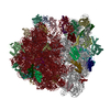
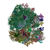
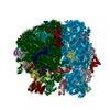
- PDB-7s1g: wild-type Escherichia coli stalled ribosome with antibiotic linezolid -
+ Open data

- Basic information
| Entry | Database: PDB / ID: 7s1g | ||||||||||||||||||||||||||||||||||||
|---|---|---|---|---|---|---|---|---|---|---|---|---|---|---|---|---|---|---|---|---|---|---|---|---|---|---|---|---|---|---|---|---|---|---|---|---|---|
| Title | wild-type Escherichia coli stalled ribosome with antibiotic linezolid | ||||||||||||||||||||||||||||||||||||
 Components Components |
| ||||||||||||||||||||||||||||||||||||
 Keywords Keywords | RIBOSOME/ANTIBIOTIC / Escherichia coli stalled ribosome /  oxazolidinone / linezolid / RIBOSOME-ANTIBIOTIC complex oxazolidinone / linezolid / RIBOSOME-ANTIBIOTIC complex | ||||||||||||||||||||||||||||||||||||
| Function / homology |  Function and homology information Function and homology informationpositive regulation of ribosome biogenesis / DnaA-L2 complex / negative regulation of DNA-templated DNA replication initiation / assembly of large subunit precursor of preribosome / cytosolic ribosome assembly / regulation of cell growth / mRNA 5'-UTR binding / ribosomal large subunit assembly / small ribosomal subunit rRNA binding / ribosomal small subunit assembly ...positive regulation of ribosome biogenesis / DnaA-L2 complex / negative regulation of DNA-templated DNA replication initiation / assembly of large subunit precursor of preribosome / cytosolic ribosome assembly / regulation of cell growth / mRNA 5'-UTR binding / ribosomal large subunit assembly / small ribosomal subunit rRNA binding / ribosomal small subunit assembly / cytosolic small ribosomal subunit / ribosome binding / large ribosomal subunit / small ribosomal subunit / cytoplasmic translation / 5S rRNA binding / cytosolic large ribosomal subunit / transferase activity / tRNA binding / negative regulation of translation / rRNA binding / ribosome / structural constituent of ribosome / translation / ribonucleoprotein complex / mRNA binding / RNA binding / zinc ion binding / membrane / metal ion binding / cytosol / cytoplasmSimilarity search - Function | ||||||||||||||||||||||||||||||||||||
| Biological species |  Escherichia coli (E. coli) Escherichia coli (E. coli) | ||||||||||||||||||||||||||||||||||||
| Method | ELECTRON MICROSCOPY / single particle reconstruction / cryo EM / Resolution: 2.48 Å | ||||||||||||||||||||||||||||||||||||
 Authors Authors | Young, I.D. / Stojkovic, V. / Tsai, K. / Lee, D.J. / Fraser, J.S. / Galonic Fujimori, D. | ||||||||||||||||||||||||||||||||||||
| Funding support | 11items
| ||||||||||||||||||||||||||||||||||||
 Citation Citation | Journal: Nat Struct Mol Biol / Year: 2022 Title: Structural basis for context-specific inhibition of translation by oxazolidinone antibiotics. Authors: Kaitlyn Tsai / Vanja Stojković / D John Lee / Iris D Young / Teresa Szal / Dorota Klepacki / Nora Vázquez-Laslop / Alexander S Mankin / James S Fraser / Danica Galonić Fujimori /  Abstract: The antibiotic linezolid, the first clinically approved member of the oxazolidinone class, inhibits translation of bacterial ribosomes by binding to the peptidyl transferase center. Recent work has ...The antibiotic linezolid, the first clinically approved member of the oxazolidinone class, inhibits translation of bacterial ribosomes by binding to the peptidyl transferase center. Recent work has demonstrated that linezolid does not inhibit peptide bond formation at all sequences but rather acts in a context-specific manner, namely when alanine occupies the penultimate position of the nascent chain. However, the molecular basis for context-specificity has not been elucidated. Here we show that the second-generation oxazolidinone radezolid also induces stalling with a penultimate alanine, and we determine high-resolution cryo-EM structures of linezolid- and radezolid-stalled ribosome complexes to explain their mechanism of action. These structures reveal that the alanine side chain fits within a small hydrophobic crevice created by oxazolidinone, resulting in improved ribosome binding. Modification of the ribosome by the antibiotic resistance enzyme Cfr disrupts stalling due to repositioning of the modified nucleotide. Together, our findings provide molecular understanding for the context-specificity of oxazolidinones. #1: Journal: Acta Crystallogr D Struct Biol / Year: 2019 Title: Macromolecular structure determination using X-rays, neutrons and electrons: recent developments in Phenix. Authors: Dorothee Liebschner / Pavel V Afonine / Matthew L Baker / Gábor Bunkóczi / Vincent B Chen / Tristan I Croll / Bradley Hintze / Li Wei Hung / Swati Jain / Airlie J McCoy / Nigel W Moriarty ...Authors: Dorothee Liebschner / Pavel V Afonine / Matthew L Baker / Gábor Bunkóczi / Vincent B Chen / Tristan I Croll / Bradley Hintze / Li Wei Hung / Swati Jain / Airlie J McCoy / Nigel W Moriarty / Robert D Oeffner / Billy K Poon / Michael G Prisant / Randy J Read / Jane S Richardson / David C Richardson / Massimo D Sammito / Oleg V Sobolev / Duncan H Stockwell / Thomas C Terwilliger / Alexandre G Urzhumtsev / Lizbeth L Videau / Christopher J Williams / Paul D Adams /   Abstract: Diffraction (X-ray, neutron and electron) and electron cryo-microscopy are powerful methods to determine three-dimensional macromolecular structures, which are required to understand biological ...Diffraction (X-ray, neutron and electron) and electron cryo-microscopy are powerful methods to determine three-dimensional macromolecular structures, which are required to understand biological processes and to develop new therapeutics against diseases. The overall structure-solution workflow is similar for these techniques, but nuances exist because the properties of the reduced experimental data are different. Software tools for structure determination should therefore be tailored for each method. Phenix is a comprehensive software package for macromolecular structure determination that handles data from any of these techniques. Tasks performed with Phenix include data-quality assessment, map improvement, model building, the validation/rebuilding/refinement cycle and deposition. Each tool caters to the type of experimental data. The design of Phenix emphasizes the automation of procedures, where possible, to minimize repetitive and time-consuming manual tasks, while default parameters are chosen to encourage best practice. A graphical user interface provides access to many command-line features of Phenix and streamlines the transition between programs, project tracking and re-running of previous tasks. #2: Journal: Acta Crystallogr D Struct Biol / Year: 2018Title: ISOLDE: a physically realistic environment for model building into low-resolution electron-density maps. Authors: Tristan Ian Croll / Abstract: This paper introduces ISOLDE, a new software package designed to provide an intuitive environment for high-fidelity interactive remodelling/refinement of macromolecular models into electron-density ...This paper introduces ISOLDE, a new software package designed to provide an intuitive environment for high-fidelity interactive remodelling/refinement of macromolecular models into electron-density maps. ISOLDE combines interactive molecular-dynamics flexible fitting with modern molecular-graphics visualization and established structural biology libraries to provide an immersive interface wherein the model constantly acts to maintain physically realistic conformations as the user interacts with it by directly tugging atoms with a mouse or haptic interface or applying/removing restraints. In addition, common validation tasks are accelerated and visualized in real time. Using the recently described 3.8 Å resolution cryo-EM structure of the eukaryotic minichromosome maintenance (MCM) helicase complex as a case study, it is demonstrated how ISOLDE can be used alongside other modern refinement tools to avoid common pitfalls of low-resolution modelling and improve the quality of the final model. A detailed analysis of changes between the initial and final model provides a somewhat sobering insight into the dangers of relying on a small number of validation metrics to judge the quality of a low-resolution model. #3: Journal: Acta Crystallogr D Biol Crystallogr / Year: 2010 Title: Features and development of Coot. Authors: P Emsley / B Lohkamp / W G Scott / K Cowtan / Abstract: Coot is a molecular-graphics application for model building and validation of biological macromolecules. The program displays electron-density maps and atomic models and allows model manipulations ...Coot is a molecular-graphics application for model building and validation of biological macromolecules. The program displays electron-density maps and atomic models and allows model manipulations such as idealization, real-space refinement, manual rotation/translation, rigid-body fitting, ligand search, solvation, mutations, rotamers and Ramachandran idealization. Furthermore, tools are provided for model validation as well as interfaces to external programs for refinement, validation and graphics. The software is designed to be easy to learn for novice users, which is achieved by ensuring that tools for common tasks are 'discoverable' through familiar user-interface elements (menus and toolbars) or by intuitive behaviour (mouse controls). Recent developments have focused on providing tools for expert users, with customisable key bindings, extensions and an extensive scripting interface. The software is under rapid development, but has already achieved very widespread use within the crystallographic community. The current state of the software is presented, with a description of the facilities available and of some of the underlying methods employed. #4: Journal: Protein Sci / Year: 2021 Title: UCSF ChimeraX: Structure visualization for researchers, educators, and developers. Authors: Eric F Pettersen / Thomas D Goddard / Conrad C Huang / Elaine C Meng / Gregory S Couch / Tristan I Croll / John H Morris / Thomas E Ferrin / Abstract: UCSF ChimeraX is the next-generation interactive visualization program from the Resource for Biocomputing, Visualization, and Informatics (RBVI), following UCSF Chimera. ChimeraX brings (a) ...UCSF ChimeraX is the next-generation interactive visualization program from the Resource for Biocomputing, Visualization, and Informatics (RBVI), following UCSF Chimera. ChimeraX brings (a) significant performance and graphics enhancements; (b) new implementations of Chimera's most highly used tools, many with further improvements; (c) several entirely new analysis features; (d) support for new areas such as virtual reality, light-sheet microscopy, and medical imaging data; (e) major ease-of-use advances, including toolbars with icons to perform actions with a single click, basic "undo" capabilities, and more logical and consistent commands; and (f) an app store for researchers to contribute new tools. ChimeraX includes full user documentation and is free for noncommercial use, with downloads available for Windows, Linux, and macOS from https://www.rbvi.ucsf.edu/chimerax. | ||||||||||||||||||||||||||||||||||||
| History |
|
- Structure visualization
| Movie |
Movie viewer |
|---|---|
| Structure viewer | Molecule: MolmilJmol/JSmol |
- Downloads & links
-Download
| PDBx/mmCIF format | 7s1g.cif.gz | 4.9 MB | Display | PDBx/mmCIF format |
|---|---|---|---|---|
| PDB format | pdb7s1g.ent.gz | Display | PDB format | |
| PDBx/mmJSON format | 7s1g.json.gz | Tree view | PDBx/mmJSON format | |
| Others |  Other downloads Other downloads |
-Validation report
| Arichive directory | https://data.pdbj.org/pub/pdb/validation_reports/s1/7s1gftp://data.pdbj.org/pub/pdb/validation_reports/s1/7s1g | HTTPS FTP |
|---|
-Related structure data
| Related structure data | 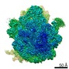 24800MC 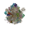 7s1hC 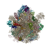 7s1iC  7s1jC 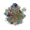 7s1kC M: map data used to model this data C: citing same article ( |
|---|---|
| Similar structure data |


-Links
 PDBj
PDBj


- Assembly
| Deposited unit | 
|
|---|---|
| 1 |
|
-Components
-30S ribosomal protein ... , 20 types, 20 molecules 123DEFGHnopqrtuvwxyz
| #1: Protein | Mass: 10455.355 Da / Num. of mol.: 1 / Source method: isolated from a natural source / Source: (natural) Escherichia coli (E. coli) / References: UniProt: D7Z9F9 |
|---|---|
| #2: Protein | Mass: 9708.464 Da / Num. of mol.: 1 / Source method: isolated from a natural source / Source: (natural) Escherichia coli (E. coli) / References: UniProt: D7ZAS2 |
| #3: Protein | Mass: 8524.039 Da / Num. of mol.: 1 / Source method: isolated from a natural source / Source: (natural) Escherichia coli (E. coli) / References: UniProt: A0A0E2L2J1 |
| #7: Protein | Mass: 26652.557 Da / Num. of mol.: 1 / Source method: isolated from a natural source / Source: (natural) Escherichia coli (E. coli) / References: UniProt: D7ZK99 |
| #8: Protein | Mass: 26031.316 Da / Num. of mol.: 1 / Source method: isolated from a natural source / Source: (natural) Escherichia coli (E. coli) / References: UniProt: B7MCS9 |
| #9: Protein | Mass: 23514.199 Da / Num. of mol.: 1 / Source method: isolated from a natural source / Source: (natural) Escherichia coli (E. coli) / References: UniProt: D7Z9H7 |
| #10: Protein | Mass: 17629.398 Da / Num. of mol.: 1 / Source method: isolated from a natural source / Source: (natural) Escherichia coli (E. coli) / References: UniProt: D7Z9H2 |
| #11: Protein | / Small ribosomal subunit protein bS6 Mass: 15727.512 Da / Num. of mol.: 1 / Source method: isolated from a natural source / Source: (natural) Escherichia coli (E. coli) / References: UniProt: P02358 |
| #43: Protein | / Small ribosomal subunit protein uS7 Mass: 20055.156 Da / Num. of mol.: 1 / Source method: isolated from a natural source / Source: (natural) Escherichia coli (E. coli) / References: UniProt: P02359 |
| #44: Protein | Mass: 14146.557 Da / Num. of mol.: 1 / Source method: isolated from a natural source / Source: (natural) Escherichia coli (E. coli) / References: UniProt: D7Z9G9 |
| #45: Protein | Mass: 14886.270 Da / Num. of mol.: 1 / Source method: isolated from a natural source / Source: (natural) Escherichia coli (E. coli) / References: UniProt: D7ZES9 |
| #46: Protein | Mass: 11755.597 Da / Num. of mol.: 1 / Source method: isolated from a natural source / Source: (natural) Escherichia coli (E. coli) / References: UniProt: D7Z9F4 |
| #47: Protein | Mass: 13870.975 Da / Num. of mol.: 1 / Source method: isolated from a natural source / Source: (natural) Escherichia coli (E. coli) / References: UniProt: B7MCR3 |
| #49: Protein | Mass: 13768.157 Da / Num. of mol.: 1 / Source method: isolated from a natural source / Source: (natural) Escherichia coli (E. coli) / References: UniProt: D7ZAN0 |
| #50: Protein | Mass: 13128.467 Da / Num. of mol.: 1 / Source method: isolated from a natural source / Source: (natural) Escherichia coli (E. coli) / References: UniProt: H4UQ02 |
| #51: Protein | Mass: 11606.560 Da / Num. of mol.: 1 / Source method: isolated from a natural source / Source: (natural) Escherichia coli (E. coli) / References: UniProt: D7Z9G8 |
| #52: Protein | Mass: 10290.816 Da / Num. of mol.: 1 / Source method: isolated from a natural source / Source: (natural) Escherichia coli (E. coli) / References: UniProt: D7ZEL4 |
| #53: Protein | Mass: 9207.572 Da / Num. of mol.: 1 / Source method: isolated from a natural source / Source: (natural) Escherichia coli (E. coli) / References: UniProt: B7MIU7 |
| #54: Protein | Mass: 9724.491 Da / Num. of mol.: 1 / Source method: isolated from a natural source / Source: (natural) Escherichia coli (E. coli) / References: UniProt: D7Z9G4 |
| #55: Protein | Mass: 9005.472 Da / Num. of mol.: 1 / Source method: isolated from a natural source / Source: (natural) Escherichia coli (E. coli) / References: UniProt: A0A0E2KXL3 |
-RNA chain , 5 types, 5 molecules 4ACIJ
| #4: RNA chain | Messenger RNA Mass: 4737.857 Da / Num. of mol.: 1 / Source method: isolated from a natural source / Source: (natural) Escherichia coli (E. coli) |
|---|---|
| #5: RNA chain | Mass: 24509.562 Da / Num. of mol.: 1 / Source method: isolated from a natural source / Source: (natural) Escherichia coli (E. coli) / References: GenBank: 1841332652 |
| #6: RNA chain | Mass: 499054.625 Da / Num. of mol.: 1 / Source method: isolated from a natural source / Source: (natural) Escherichia coli (E. coli) / References: GenBank: 1758835854 |
| #12: RNA chain | 23S ribosomal RNA Mass: 941811.562 Da / Num. of mol.: 1 / Source method: isolated from a natural source / Source: (natural) Escherichia coli (E. coli) |
| #13: RNA chain | 5S ribosomal RNA Mass: 38790.090 Da / Num. of mol.: 1 / Source method: isolated from a natural source / Source: (natural) Escherichia coli (E. coli) / References: GenBank: 1273279017 |
+50S ribosomal protein ... , 29 types, 29 molecules KLMNOPQRSTUVWXYZabcdefghijklm
-Protein/peptide , 1 types, 1 molecules s
| #48: Protein/peptide | Mass: 643.816 Da / Num. of mol.: 1 / Source method: isolated from a natural source / Source: (natural) Escherichia coli (E. coli) |
|---|
-Non-polymers , 4 types, 241 molecules 


| #56: Chemical | ChemComp-MG /  Mass: 24.305 Da / Num. of mol.: 196 / Source method: obtained synthetically / Formula: Mg Mass: 24.305 Da / Num. of mol.: 196 / Source method: obtained synthetically / Formula: Mg#57: Chemical | ChemComp-ZLD / | Linezolid Mass: 337.346 Da / Num. of mol.: 1 / Source method: obtained synthetically / Formula: C16H20FN3O4 / Feature type: SUBJECT OF INVESTIGATION / Comment: antibiotic*YM Mass: 337.346 Da / Num. of mol.: 1 / Source method: obtained synthetically / Formula: C16H20FN3O4 / Feature type: SUBJECT OF INVESTIGATION / Comment: antibiotic*YM#58: Chemical |  Mass: 65.409 Da / Num. of mol.: 2 / Source method: obtained synthetically / Formula: Zn Mass: 65.409 Da / Num. of mol.: 2 / Source method: obtained synthetically / Formula: Zn#59: Water | ChemComp-HOH / | WaterMass: 18.015 Da / Num. of mol.: 42 / Source method: isolated from a natural source / Formula: H2O |
|---|
-Details
| Has ligand of interest | Y |
|---|
-Experimental details
-Experiment
| Experiment | Method: ELECTRON MICROSCOPY |
|---|---|
| EM experiment | Aggregation state: PARTICLE / 3D reconstruction method: single particle reconstruction |
- Sample preparation
| Component | Name: wild-type Escherichia coli stalled ribosome with antibiotic linezolid Type: RIBOSOME / Entity ID: #1-#55 / Source: NATURAL |
|---|---|
| Molecular weight | Value: 2.154 MDa / Experimental value: NO |
| Source (natural) | Organism: Escherichia coli (E. coli) |
| Buffer solution | pH: 7.5 |
| Specimen | Embedding applied: NO / Shadowing applied: NO / Staining applied: NO / Vitrification applied: YES |
| Specimen support | Details: 15 mA / Grid material: COPPER / Grid mesh size: 300 divisions/in. / Grid type: Quantifoil R1.2/1.3 |
| Vitrification | Instrument: FEI VITROBOT MARK IV / Cryogen name: ETHANE / Humidity: 95 % / Chamber temperature: 283.2 K |
- Electron microscopy imaging
| Experimental equipment |  Model: Titan Krios / Image courtesy: FEI Company |
|---|---|
| Microscopy | Model: FEI TITAN KRIOS |
| Electron gun | Electron source: FIELD EMISSION GUN / Accelerating voltage: 300 kV / Illumination mode: FLOOD BEAM |
| Electron lens | Mode: BRIGHT FIELDBright-field microscopy |
| Specimen holder | Cryogen: NITROGEN / Specimen holder model: FEI TITAN KRIOS AUTOGRID HOLDER |
| Image recording | Electron dose: 67.8 e/Å2 / Film or detector model: GATAN K3 (6k x 4k) / Num. of grids imaged: 1 / Num. of real images: 3644 |
- Processing
| EM software |
| ||||||||||||||||||||||||||||||||||||||||||||||||||||||||
|---|---|---|---|---|---|---|---|---|---|---|---|---|---|---|---|---|---|---|---|---|---|---|---|---|---|---|---|---|---|---|---|---|---|---|---|---|---|---|---|---|---|---|---|---|---|---|---|---|---|---|---|---|---|---|---|---|---|
| CTF correction | Type: PHASE FLIPPING AND AMPLITUDE CORRECTION | ||||||||||||||||||||||||||||||||||||||||||||||||||||||||
| Particle selection | Num. of particles selected: 332502 | ||||||||||||||||||||||||||||||||||||||||||||||||||||||||
| Symmetry | Point symmetry: C1 (asymmetric) | ||||||||||||||||||||||||||||||||||||||||||||||||||||||||
| 3D reconstruction | Resolution: 2.48 Å / Resolution method: FSC 0.143 CUT-OFF / Num. of particles: 176779 / Num. of class averages: 1 / Symmetry type: POINT | ||||||||||||||||||||||||||||||||||||||||||||||||||||||||
| Atomic model building | Protocol: FLEXIBLE FIT / Space: REAL |
