 Movie
Movie Controller

[English] 日本語
 Yorodumi
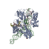
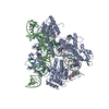
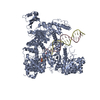
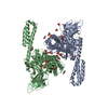
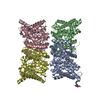
- PDB-7pw6: Human SMG1-8-9 kinase complex bound to a SMG1 inhibitor - SMG1 body -
+ Open data

- Basic information
| Entry | Database: PDB / ID: 7pw6 | ||||||
|---|---|---|---|---|---|---|---|
| Title | Human SMG1-8-9 kinase complex bound to a SMG1 inhibitor - SMG1 body | ||||||
 Components Components | Serine/threonine-protein kinase SMG1,Serine/threonine-protein kinase SMG1,Serine/threonine-protein kinase SMG1,Serine/threonine-protein kinase SMG1,Serine/threonine-protein kinase SMG1,Serine/threonine-protein kinase SMG1,Serine/threonine-protein kinase SMG1 | ||||||
 Keywords Keywords |  TRANSFERASE / Complex TRANSFERASE / Complex | ||||||
| Function / homology |  Function and homology information Function and homology informationdiacylglycerol-dependent serine/threonine kinase activity / chromatoid body / regulation of telomere maintenance / nuclear-transcribed mRNA catabolic process, nonsense-mediated decay / telomeric DNA binding / phosphatidylinositol phosphate biosynthetic process / Nonsense Mediated Decay (NMD) enhanced by the Exon Junction Complex (EJC) / mRNA export from nucleus / peptidyl-serine phosphorylation / protein autophosphorylation ...diacylglycerol-dependent serine/threonine kinase activity / chromatoid body / regulation of telomere maintenance / nuclear-transcribed mRNA catabolic process, nonsense-mediated decay / telomeric DNA binding / phosphatidylinositol phosphate biosynthetic process / Nonsense Mediated Decay (NMD) enhanced by the Exon Junction Complex (EJC) / mRNA export from nucleus / peptidyl-serine phosphorylation / protein autophosphorylation / non-specific serine/threonine protein kinase / protein kinase activity / protein serine kinase activity / DNA repair / protein serine/threonine kinase activity / DNA damage response / RNA binding / nucleoplasm / ATP binding / metal ion binding / nucleus / cytosol / cytoplasmSimilarity search - Function | ||||||
| Biological species |  Homo sapiens (human) Homo sapiens (human) | ||||||
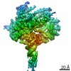
| Method | ELECTRON MICROSCOPY / single particle reconstruction / cryo EM / Resolution: 3.05 Å | ||||||
 Authors Authors | Langer, L.M. / Conti, E. | ||||||
| Funding support |  Germany, 1items Germany, 1items
| ||||||
 Citation Citation | Journal: Elife / Year: 2021 Title: Cryo-EM reconstructions of inhibitor-bound SMG1 kinase reveal an autoinhibitory state dependent on SMG8. Authors: Lukas M Langer / Fabien Bonneau / Yair Gat / Elena Conti / Abstract: The PI3K-related kinase (PIKK) SMG1 monitors the progression of metazoan nonsense-mediated mRNA decay (NMD) by phosphorylating the RNA helicase UPF1. Previous work has shown that the activity of SMG1 ...The PI3K-related kinase (PIKK) SMG1 monitors the progression of metazoan nonsense-mediated mRNA decay (NMD) by phosphorylating the RNA helicase UPF1. Previous work has shown that the activity of SMG1 is impaired by small molecule inhibitors, is reduced by the SMG1 interactors SMG8 and SMG9, and is downregulated by the so-called SMG1 insertion domain. However, the molecular basis for this complex regulatory network has remained elusive. Here, we present cryo-electron microscopy reconstructions of human SMG1-9 and SMG1-8-9 complexes bound to either a SMG1 inhibitor or a non-hydrolyzable ATP analog at overall resolutions ranging from 2.8 to 3.6 Å. These structures reveal the basis with which a small molecule inhibitor preferentially targets SMG1 over other PIKKs. By comparison with our previously reported substrate-bound structure (Langer et al.,2020), we show that the SMG1 insertion domain can exert an autoinhibitory function by directly blocking the substrate-binding path as well as overall access to the SMG1 kinase active site. Together with biochemical analysis, our data indicate that SMG1 autoinhibition is stabilized by the presence of SMG8. Our results explain the specific inhibition of SMG1 by an ATP-competitive small molecule, provide insights into regulation of its kinase activity within the NMD pathway, and expand the understanding of PIKK regulatory mechanisms in general. | ||||||
| History |
|
- Structure visualization
| Movie |
Movie viewer |
|---|---|
| Structure viewer | Molecule: MolmilJmol/JSmol |
- Downloads & links
-Download
| PDBx/mmCIF format | 7pw6.cif.gz | 278.2 KB | Display | PDBx/mmCIF format |
|---|---|---|---|---|
| PDB format | pdb7pw6.ent.gz | 202.6 KB | Display | PDB format |
| PDBx/mmJSON format | 7pw6.json.gz | Tree view | PDBx/mmJSON format | |
| Others |  Other downloads Other downloads |
-Validation report
| Arichive directory | https://data.pdbj.org/pub/pdb/validation_reports/pw/7pw6ftp://data.pdbj.org/pub/pdb/validation_reports/pw/7pw6 | HTTPS FTP |
|---|
-Related structure data
| Related structure data |  13676MC  7pw4C  7pw5C  7pw7C  7pw8C  7pw9C M: map data used to model this data C: citing same article ( |
|---|---|
| Similar structure data |


-Links
 PDBj
PDBj


- Assembly
| Deposited unit | 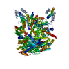
|
|---|---|
| 1 |
|
-Components
| #1: Protein | Mass: 320901.969 Da / Num. of mol.: 1 Source method: isolated from a genetically manipulated source Source: (gene. exp.) Homo sapiens (human) / Gene: SMG1, ATX, KIAA0421, LIP / Production host: Homo sapiens (human)References: UniProt: Q96Q15, non-specific serine/threonine protein kinase |
|---|---|
| #2: Chemical | ChemComp-IHP / Phytic acid  Mass: 660.035 Da / Num. of mol.: 1 / Source method: obtained synthetically / Formula: C6H18O24P6 Mass: 660.035 Da / Num. of mol.: 1 / Source method: obtained synthetically / Formula: C6H18O24P6 |
| #3: Chemical | ChemComp-88C / 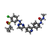  Mass: 566.074 Da / Num. of mol.: 1 / Source method: obtained synthetically / Formula: C27H28ClN7O3S / Feature type: SUBJECT OF INVESTIGATION Mass: 566.074 Da / Num. of mol.: 1 / Source method: obtained synthetically / Formula: C27H28ClN7O3S / Feature type: SUBJECT OF INVESTIGATION |
| Has ligand of interest | Y |
-Experimental details
-Experiment
| Experiment | Method: ELECTRON MICROSCOPY |
|---|---|
| EM experiment | Aggregation state: PARTICLE / 3D reconstruction method: single particle reconstruction |
- Sample preparation
| Component | Name: SMG1-SMG8-SMG9 kinase complex bound to a SMG1 inhibitor Type: COMPLEX / Entity ID: #1 / Source: RECOMBINANT |
|---|---|
| Molecular weight | Value: 0.597 MDa / Experimental value: NO |
| Source (natural) | Organism: Homo sapiens (human) |
| Source (recombinant) | Organism: Homo sapiens (human) |
| Buffer solution | pH: 7.4 |
| Specimen | Embedding applied: NO / Shadowing applied: NO / Staining applied: NO / Vitrification applied: YES |
| Vitrification | Cryogen name: ETHANE-PROPANE |
- Electron microscopy imaging
| Experimental equipment |  Model: Titan Krios / Image courtesy: FEI Company |
|---|---|
| Microscopy | Model: FEI TITAN KRIOS |
| Electron gun | Electron source: FIELD EMISSION GUN / Accelerating voltage: 300 kV / Illumination mode: FLOOD BEAM |
| Electron lens | Mode: BRIGHT FIELDBright-field microscopy |
| Image recording | Electron dose: 89.32 e/Å2 / Film or detector model: GATAN K3 (6k x 4k) |
- Processing
| Software | Name: PHENIX / Version: 1.19.2_4158: / Classification: refinement | ||||||||||||||||||||||||
|---|---|---|---|---|---|---|---|---|---|---|---|---|---|---|---|---|---|---|---|---|---|---|---|---|---|
| CTF correction | Type: PHASE FLIPPING AND AMPLITUDE CORRECTION | ||||||||||||||||||||||||
| 3D reconstruction | Resolution: 3.05 Å / Resolution method: FSC 0.143 CUT-OFF / Num. of particles: 228291 / Symmetry type: POINT | ||||||||||||||||||||||||
| Refine LS restraints |
|
