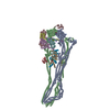
 Movie
Movie Controller

[English] 日本語
 Yorodumi
- PDB-7nyz: Cryo-EM structure of the MukBEF-MatP-DNA monomer (partially open ... -
+ Open data

- Basic information
| Entry | Database: PDB / ID: 7nyz | ||||||
|---|---|---|---|---|---|---|---|
| Title | Cryo-EM structure of the MukBEF-MatP-DNA monomer (partially open conformation) | ||||||
 Components Components |
| ||||||
 Keywords Keywords |  DNA BINDING PROTEIN / SMC-kleisin complex / ATPase DNA BINDING PROTEIN / SMC-kleisin complex / ATPase | ||||||
| Function / homology |  Function and homology informationnucleoid / chromosome condensation / acyl carrier activity / chromosome segregation / DNA replication / sequence-specific DNA binding / cell cycle / cell division / calcium ion binding / regulation of DNA-templated transcription ...nucleoid / chromosome condensation / acyl carrier activity / chromosome segregation / DNA replication / sequence-specific DNA binding / cell cycle / cell division / calcium ion binding / regulation of DNA-templated transcription / DNA binding / ATP binding / cytoplasm Function and homology informationnucleoid / chromosome condensation / acyl carrier activity / chromosome segregation / DNA replication / sequence-specific DNA binding / cell cycle / cell division / calcium ion binding / regulation of DNA-templated transcription ...nucleoid / chromosome condensation / acyl carrier activity / chromosome segregation / DNA replication / sequence-specific DNA binding / cell cycle / cell division / calcium ion binding / regulation of DNA-templated transcription / DNA binding / ATP binding / cytoplasmSimilarity search - Function | ||||||
| Biological species |  Photorhabdus thracensis (bacteria)Escherichia coli BL21 (bacteria) Photorhabdus thracensis (bacteria)Escherichia coli BL21 (bacteria) | ||||||
| Method | ELECTRON MICROSCOPY / single particle reconstruction / cryo EM / Resolution: 6.5 Å | ||||||
 Authors Authors | Buermann, F. / Lowe, J. | ||||||
 Citation Citation | Journal: Mol Cell / Year: 2021 Title: Cryo-EM structure of MukBEF reveals DNA loop entrapment at chromosomal unloading sites. Authors: Frank Bürmann / Louise F H Funke / Jason W Chin / Jan Löwe /  Abstract: The ring-like structural maintenance of chromosomes (SMC) complex MukBEF folds the genome of Escherichia coli and related bacteria into large loops, presumably by active DNA loop extrusion. MukBEF ...The ring-like structural maintenance of chromosomes (SMC) complex MukBEF folds the genome of Escherichia coli and related bacteria into large loops, presumably by active DNA loop extrusion. MukBEF activity within the replication terminus macrodomain is suppressed by the sequence-specific unloader MatP. Here, we present the complete atomic structure of MukBEF in complex with MatP and DNA as determined by electron cryomicroscopy (cryo-EM). The complex binds two distinct DNA double helices corresponding to the arms of a plectonemic loop. MatP-bound DNA threads through the MukBEF ring, while the second DNA is clamped by the kleisin MukF, MukE, and the MukB ATPase heads. Combinatorial cysteine cross-linking confirms this topology of DNA loop entrapment in vivo. Our findings illuminate how a class of near-ubiquitous DNA organizers with important roles in genome maintenance interacts with the bacterial chromosome. #1: Journal: Biorxiv / Year: 2021Title: DNA entrapment revealed by the structure of bacterial condensin MukBEF Authors: Buermann, F. / Funke, L.F.H. / Chin, J.W. / Lowe, J. | ||||||
| History |
|
- Structure visualization
| Movie |
Movie viewer |
|---|---|
| Structure viewer | Molecule: MolmilJmol/JSmol |
- Downloads & links
-Download
| PDBx/mmCIF format | 7nyz.cif.gz | 1.4 MB | Display | PDBx/mmCIF format |
|---|---|---|---|---|
| PDB format | pdb7nyz.ent.gz | 1.2 MB | Display | PDB format |
| PDBx/mmJSON format | 7nyz.json.gz | Tree view | PDBx/mmJSON format | |
| Others |  Other downloads Other downloads |
-Validation report
| Arichive directory | https://data.pdbj.org/pub/pdb/validation_reports/ny/7nyzftp://data.pdbj.org/pub/pdb/validation_reports/ny/7nyz | HTTPS FTP |
|---|
-Related structure data
| Related structure data |  12659MC  7nywC  7nyxC 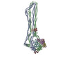 7nyyC  7nz0C 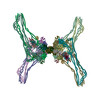 7nz2C 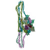 7nz3C 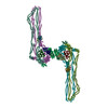 7nz4C M: map data used to model this data C: citing same article ( |
|---|---|
| Similar structure data | |
| EM raw data | EMPIAR-10755 (Title: MukBEF(E1407Q)-MatP-DNA in the presence of ATP / Data size: 8.5 TB Data #1: Unaligned multi-frame images of MukBEF(E1407Q)-MatP-DNA in the presence of ATP - Dataset 1 [micrographs - multiframe] Data #2: Unaligned multi-frame images of MukBEF(E1407Q)-MatP-DNA in the presence of ATP - Dataset 2 [micrographs - multiframe] Data #3: Unaligned multi-frame images of MukBEF(E1407Q)-MatP-DNA in the presence of ATP - Dataset 3, grid 1 [micrographs - multiframe] Data #4: Unaligned multi-frame images of MukBEF(E1407Q)-MatP-DNA in the presence of ATP - Dataset 3, grid 2 [micrographs - multiframe] Data #5: Unaligned multi-frame images of MukBEF(E1407Q)-MatP-DNA in the presence of ATP - Dataset 3, grid 3 [micrographs - multiframe]) |


-Links
 PDBj
PDBj


- Assembly
| Deposited unit | 
|
|---|---|
| 1 |
|
-Components
-Chromosome partition protein ... , 3 types, 6 molecules ABCDEF
| #1: Protein | Mass: 170240.188 Da / Num. of mol.: 2 / Mutation: E1407Q Source method: isolated from a genetically manipulated source Source: (gene. exp.) Photorhabdus thracensis (bacteria) / Gene: mukB, VY86_15870 / Production host: Escherichia coli BL21(DE3) (bacteria) / References: UniProt: A0A0F7LRY2#2: Protein | Mass: 50193.305 Da / Num. of mol.: 2 Source method: isolated from a genetically manipulated source Source: (gene. exp.) Photorhabdus thracensis (bacteria) / Gene: mukF, VY86_15860 / Production host: Escherichia coli BL21(DE3) (bacteria) / References: UniProt: A0A0F7LMQ4#3: Protein | Mass: 27423.848 Da / Num. of mol.: 2 Source method: isolated from a genetically manipulated source Source: (gene. exp.) Photorhabdus thracensis (bacteria) / Gene: mukE, VY86_15865 / Production host: Escherichia coli BL21(DE3) (bacteria) / References: UniProt: A0A0F7LPV6 |
|---|
-Protein , 2 types, 4 molecules GHIJ
| #4: Protein | / ACP Mass: 8645.460 Da / Num. of mol.: 2 / Source method: isolated from a natural source / Source: (natural) Escherichia coli BL21(DE3) (bacteria) / References: UniProt: A0A6D2XA84#5: Protein | Mass: 18032.613 Da / Num. of mol.: 2 Source method: isolated from a genetically manipulated source Source: (gene. exp.) Photorhabdus thracensis (bacteria) / Gene: matP, VY86_22090 / Production host: Escherichia coli BL21(DE3) (bacteria) / References: UniProt: A0A0F7LUV5 |
|---|
-MatS2 DNA 80 b, oligo ... , 2 types, 2 molecules KL
| #6: DNA chain | Mass: 24625.713 Da / Num. of mol.: 1 / Source method: obtained synthetically / Details: Synthetic sequence / Source: (synth.) Photorhabdus thracensis (bacteria) |
|---|---|
| #7: DNA chain | Mass: 24715.855 Da / Num. of mol.: 1 / Source method: obtained synthetically / Details: Synthetic sequence / Source: (synth.) Photorhabdus thracensis (bacteria) |
-DNA chain , 1 types, 2 molecules MN
| #8: DNA chain | Mass: 9216.037 Da / Num. of mol.: 2 / Source method: obtained synthetically / Details: Synthetic sequence / Source: (synth.) Photorhabdus thracensis (bacteria) |
|---|
-Non-polymers , 3 types, 6 molecules 

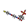
| #9: Chemical |  Mass: 24.305 Da / Num. of mol.: 2 / Source method: obtained synthetically / Formula: Mg Mass: 24.305 Da / Num. of mol.: 2 / Source method: obtained synthetically / Formula: Mg#10: Chemical | Adenosine triphosphate Mass: 507.181 Da / Num. of mol.: 2 / Source method: obtained synthetically / Formula: C10H16N5O13P3 / Comment: ATP, energy-carrying molecule*YM Mass: 507.181 Da / Num. of mol.: 2 / Source method: obtained synthetically / Formula: C10H16N5O13P3 / Comment: ATP, energy-carrying molecule*YM#11: Chemical | Phosphopantetheine Mass: 358.348 Da / Num. of mol.: 2 / Source method: obtained synthetically / Formula: C11H23N2O7PS Mass: 358.348 Da / Num. of mol.: 2 / Source method: obtained synthetically / Formula: C11H23N2O7PS |
|---|
-Details
| Has ligand of interest | N |
|---|
-Experimental details
-Experiment
| Experiment | Method: ELECTRON MICROSCOPY |
|---|---|
| EM experiment | Aggregation state: PARTICLE / 3D reconstruction method: single particle reconstruction |
- Sample preparation
| Component |
| ||||||||||||||||||||||||||||||
|---|---|---|---|---|---|---|---|---|---|---|---|---|---|---|---|---|---|---|---|---|---|---|---|---|---|---|---|---|---|---|---|
| Molecular weight | Experimental value: NO | ||||||||||||||||||||||||||||||
| Source (natural) |
| ||||||||||||||||||||||||||||||
| Source (recombinant) |
| ||||||||||||||||||||||||||||||
| Buffer solution | pH: 7.3 | ||||||||||||||||||||||||||||||
| Specimen | Embedding applied: NO / Shadowing applied: NO / Staining applied: NO / Vitrification applied: YES | ||||||||||||||||||||||||||||||
| Vitrification | Cryogen name: ETHANE |
- Electron microscopy imaging
| Experimental equipment |  Model: Titan Krios / Image courtesy: FEI Company |
|---|---|
| Microscopy | Model: FEI TITAN KRIOS |
| Electron gun | Electron source: FIELD EMISSION GUN / Accelerating voltage: 300 kV / Illumination mode: OTHER |
| Electron lens | Mode: BRIGHT FIELDBright-field microscopy |
| Image recording | Electron dose: 40 e/Å2 / Film or detector model: GATAN K3 (6k x 4k) |
- Processing
| Software | Name: UCSF ChimeraX / Version: 1.1/v9 / Classification: model building / URL: https://www.rbvi.ucsf.edu/chimerax/ / Os: Windows / Type: package |
|---|---|
| CTF correction | Type: PHASE FLIPPING AND AMPLITUDE CORRECTION |
| 3D reconstruction | Resolution: 6.5 Å / Resolution method: FSC 0.143 CUT-OFF / Num. of particles: 41109 / Symmetry type: POINT |
