 Movie
Movie Controller


+ Open data

- Basic information
| Entry | Database: PDB / ID: 7ks3 | ||||||
|---|---|---|---|---|---|---|---|
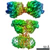
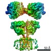
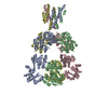
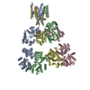
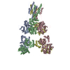
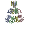
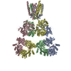
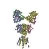
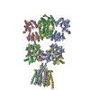
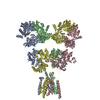
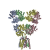
| Title | GluK2/K5 with L-Glu | ||||||
 Components Components |
| ||||||
 Keywords Keywords |  SIGNALING PROTEIN / Kainate receptor / ionotropic glutamate receptor / membrane protein / ligand-gated ion channel SIGNALING PROTEIN / Kainate receptor / ionotropic glutamate receptor / membrane protein / ligand-gated ion channel | ||||||
| Function / homology |  Function and homology information Function and homology informationregulation of synaptic vesicle fusion to presynaptic active zone membrane / protein retention in ER lumen / mossy fiber rosette / detection of cold stimulus involved in thermoception / Activation of Na-permeable kainate receptors / kainate selective glutamate receptor complex / Activation of Ca-permeable Kainate Receptor / negative regulation of synaptic transmission, glutamatergic / regulation of short-term neuronal synaptic plasticity / inhibitory postsynaptic potential ...regulation of synaptic vesicle fusion to presynaptic active zone membrane / protein retention in ER lumen / mossy fiber rosette / detection of cold stimulus involved in thermoception / Activation of Na-permeable kainate receptors / kainate selective glutamate receptor complex / Activation of Ca-permeable Kainate Receptor / negative regulation of synaptic transmission, glutamatergic / regulation of short-term neuronal synaptic plasticity / inhibitory postsynaptic potential / glutamate receptor activity / ubiquitin conjugating enzyme binding / receptor clustering / modulation of excitatory postsynaptic potential / regulation of JNK cascade / kainate selective glutamate receptor activity / ionotropic glutamate receptor complex / extracellularly glutamate-gated ion channel activity / neuronal action potential / behavioral fear response / positive regulation of synaptic transmission / glutamate-gated receptor activity / ligand-gated monoatomic ion channel activity involved in regulation of presynaptic membrane potential / presynaptic modulation of chemical synaptic transmission / excitatory postsynaptic potential / dendrite cytoplasm / hippocampal mossy fiber to CA3 synapse / bioluminescence / SNARE binding / generation of precursor metabolites and energy / synaptic transmission, glutamatergic / regulation of membrane potential / transmitter-gated monoatomic ion channel activity involved in regulation of postsynaptic membrane potential / establishment of localization in cell / PDZ domain binding / cellular response to glucose stimulus / postsynaptic density membrane / regulation of long-term neuronal synaptic plasticity / modulation of chemical synaptic transmission / terminal bouton / SH3 domain binding / intracellular calcium ion homeostasis / positive regulation of neuron apoptotic process / presynaptic membrane / scaffold protein binding / chemical synaptic transmission / postsynaptic membrane / perikaryon / neuron apoptotic process / negative regulation of neuron apoptotic process / postsynaptic density / axon / neuronal cell body / glutamatergic synapse / synapse / ubiquitin protein ligase binding / dendrite / endoplasmic reticulum / membrane / identical protein binding / plasma membraneSimilarity search - Function | ||||||
| Biological species |  Rattus norvegicus (Norway rat) Rattus norvegicus (Norway rat)  Aequorea victoria (jellyfish) Aequorea victoria (jellyfish) | ||||||
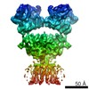
| Method | ELECTRON MICROSCOPY / single particle reconstruction / cryo EM / Resolution: 5.8 Å | ||||||
 Authors Authors | Khanra, N. / Brown, P.M.G.E. / Perozzo, A.M. / Bowie, D. / Meyerson, J.R. | ||||||
 Citation Citation | Journal: Elife / Year: 2021 Title: Architecture and structural dynamics of the heteromeric GluK2/K5 kainate receptor. Authors: Nandish Khanra / Patricia Mge Brown / Amanda M Perozzo / Derek Bowie / Joel R Meyerson /   Abstract: Kainate receptors (KARs) are L-glutamate-gated ion channels that regulate synaptic transmission and modulate neuronal circuits. KARs have strict assembly rules and primarily function as heteromeric ...Kainate receptors (KARs) are L-glutamate-gated ion channels that regulate synaptic transmission and modulate neuronal circuits. KARs have strict assembly rules and primarily function as heteromeric receptors in the brain. A longstanding question is how KAR heteromer subunits organize and coordinate together to fulfill their signature physiological roles. Here we report structures of the GluK2/GluK5 heteromer in apo, antagonist-bound, and desensitized states. The receptor assembles with two copies of each subunit, ligand binding domains arranged as two heterodimers and GluK5 subunits proximal to the channel. Strikingly, during desensitization, GluK2, but not GluK5, subunits undergo major structural rearrangements to facilitate channel closure. We show how the large conformational differences between antagonist-bound and desensitized states are mediated by the linkers connecting the pore helices to the ligand binding domains. This work presents the first KAR heteromer structure, reveals how its subunits are organized, and resolves how the heteromer can accommodate functionally distinct closed channel structures. | ||||||
| History |
|
- Structure visualization
| Movie |
Movie viewer |
|---|---|
| Structure viewer | Molecule: MolmilJmol/JSmol |
- Downloads & links
-Download
| PDBx/mmCIF format | 7ks3.cif.gz | 477 KB | Display | PDBx/mmCIF format |
|---|---|---|---|---|
| PDB format | pdb7ks3.ent.gz | 369.3 KB | Display | PDB format |
| PDBx/mmJSON format | 7ks3.json.gz | Tree view | PDBx/mmJSON format | |
| Others |  Other downloads Other downloads |
-Validation report
| Arichive directory | https://data.pdbj.org/pub/pdb/validation_reports/ks/7ks3ftp://data.pdbj.org/pub/pdb/validation_reports/ks/7ks3 | HTTPS FTP |
|---|
-Related structure data
| Related structure data | 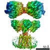 23015MC  7ks0C M: map data used to model this data C: citing same article ( |
|---|---|
| Similar structure data | |
| EM raw data | EMPIAR-10660 (Title: GluK2/K5 with L-Glu / Data size: 3.4 TB / Data #1: GluK2/K5 with L-Glu [micrographs - multiframe] / Data #2: GluK2/K5 with L-Glu [micrographs - multiframe] Data #3: GluK2/K5 with L-Glu [picked particles - single frame - processed] Data #4: GluK2/K5 with L-Glu [picked particles - single frame - processed]) |
-Links
 PDBj
PDBj


- Assembly
| Deposited unit | 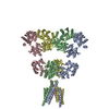
|
|---|---|
| 1 |
|
-Components
| #1: Protein | Mass: 123676.812 Da / Num. of mol.: 2 Source method: isolated from a genetically manipulated source Source: (gene. exp.) Rattus norvegicus (Norway rat), (gene. exp.) Aequorea victoria (jellyfish)Gene: Grik5, GFP / Cell line (production host): HEK293S GnTI- / Production host:  Homo sapiens (human) / References: UniProt: Q63273, UniProt: P42212 Homo sapiens (human) / References: UniProt: Q63273, UniProt: P42212#2: Protein | Mass: 105981.617 Da / Num. of mol.: 2 Source method: isolated from a genetically manipulated source Source: (gene. exp.) Rattus norvegicus (Norway rat) / Gene: Grik2, Glur6 / Cell line (production host): HEK293S GnTI- / Production host: Homo sapiens (human) / References: UniProt: P42260 |
|---|
-Experimental details
-Experiment
| Experiment | Method: ELECTRON MICROSCOPY |
|---|---|
| EM experiment | Aggregation state: PARTICLE / 3D reconstruction method: single particle reconstruction |
- Sample preparation
| Component | Name: GluK2/K5 with L-Glu / Type: COMPLEX / Entity ID: all / Source: RECOMBINANT | |||||||||||||||
|---|---|---|---|---|---|---|---|---|---|---|---|---|---|---|---|---|
| Molecular weight | Experimental value: NO | |||||||||||||||
| Source (natural) | Organism: Rattus norvegicus (Norway rat) | |||||||||||||||
| Source (recombinant) | Organism: Homo sapiens (human) / Cell: HEK293S GnTI- | |||||||||||||||
| Buffer solution | pH: 8 | |||||||||||||||
| Buffer component |
| |||||||||||||||
| Specimen | Conc.: 3.7 mg/ml / Embedding applied: NO / Shadowing applied: NO / Staining applied: NO / Vitrification applied: YES / Details: 1 mM L-Glu | |||||||||||||||
| Vitrification | Instrument: FEI VITROBOT MARK IV / Cryogen name: ETHANE / Humidity: 100 % / Chamber temperature: 295 K |
- Electron microscopy imaging
| Experimental equipment |  Model: Talos Arctica / Image courtesy: FEI Company |
|---|---|
| Microscopy | Model: FEI TECNAI ARCTICA |
| Electron gun | Electron source: FIELD EMISSION GUN / Accelerating voltage: 200 kV / Illumination mode: OTHER |
| Electron lens | Mode: BRIGHT FIELDBright-field microscopy |
| Image recording | Electron dose: 51.5 e/Å2 / Film or detector model: GATAN K3 (6k x 4k) |
- Processing
| EM software | Name: Leginon / Category: image acquisition |
|---|---|
| CTF correction | Type: PHASE FLIPPING AND AMPLITUDE CORRECTION |
| 3D reconstruction | Resolution: 5.8 Å / Resolution method: FSC 0.143 CUT-OFF / Num. of particles: 573403 Details: Number of particles for ATD map: 241849 Number of particles for LBD-TMD map: 140028 Symmetry type: POINT |
