 Movie
Movie Controller


+ Open data

- Basic information
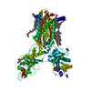
| Entry | Database: PDB / ID: 7e2h | |||||||||
|---|---|---|---|---|---|---|---|---|---|---|
| Title | Cryo-EM structure of hDisp1NNN-3C-Cleavage | |||||||||
 Components Components | (Protein dispatched homolog 1) x 2 | |||||||||
 Keywords Keywords | LIPID TRANSPORT /  membrane protein membrane protein | |||||||||
| Function / homology |  Function and homology information Function and homology informationpatched ligand maturation / diaphragm development / embryonic pattern specification / peptide transport / peptide transmembrane transporter activity / dorsal/ventral pattern formation / determination of left/right symmetry / smoothened signaling pathway / regulation of protein secretion / protein homotrimerization ...patched ligand maturation / diaphragm development / embryonic pattern specification / peptide transport / peptide transmembrane transporter activity / dorsal/ventral pattern formation / determination of left/right symmetry / smoothened signaling pathway / regulation of protein secretion / protein homotrimerization / basolateral plasma membrane / membraneSimilarity search - Function | |||||||||
| Biological species |  Homo sapiens (human) Homo sapiens (human) | |||||||||
| Method | ELECTRON MICROSCOPY / single particle reconstruction / cryo EM / Resolution: 3.68 Å | |||||||||
 Authors Authors | Li, W. / Wang, L. / Gong, X. | |||||||||
| Funding support |  China, 2items China, 2items
| |||||||||
 Citation Citation | Journal: Nat Commun / Year: 2021 Title: Structural insights into proteolytic activation of the human Dispatched1 transporter for Hedgehog morphogen release. Authors: Wanqiu Li / Linlin Wang / Bradley M Wierbowski / Mo Lu / Feitong Dong / Wenchen Liu / Sisi Li / Peiyi Wang / Adrian Salic / Xin Gong /  Abstract: The membrane protein Dispatched (Disp), which belongs to the RND family of small molecule transporters, is essential for Hedgehog (Hh) signaling, by catalyzing the extracellular release of palmitate- ...The membrane protein Dispatched (Disp), which belongs to the RND family of small molecule transporters, is essential for Hedgehog (Hh) signaling, by catalyzing the extracellular release of palmitate- and cholesterol-modified Hh ligands from producing cells. Disp function requires Furin-mediated proteolytic cleavage of its extracellular domain, but how this activates Disp remains obscure. Here, we employ cryo-electron microscopy to determine atomic structures of human Disp1 (hDisp1), before and after cleavage, and in complex with lipid-modified Sonic hedgehog (Shh) ligand. These structures, together with biochemical data, reveal that proteolytic cleavage opens the extracellular domain of hDisp1, removing steric hindrance to Shh binding. Structure-guided functional experiments demonstrate the role of hDisp1-Shh interactions in ligand release. Our results clarify the mechanisms of hDisp1 activation and Shh morphogen release, and highlight how a unique proteolytic cleavage event enabled acquisition of a protein substrate by a member of a family of small molecule transporters. | |||||||||
| History |
|
- Structure visualization
| Movie |
Movie viewer |
|---|---|
| Structure viewer | Molecule: MolmilJmol/JSmol |
- Downloads & links
-Download
| PDBx/mmCIF format | 7e2h.cif.gz | 185.8 KB | Display | PDBx/mmCIF format |
|---|---|---|---|---|
| PDB format | pdb7e2h.ent.gz | 143 KB | Display | PDB format |
| PDBx/mmJSON format | 7e2h.json.gz | Tree view | PDBx/mmJSON format | |
| Others |  Other downloads Other downloads |
-Validation report
| Arichive directory | https://data.pdbj.org/pub/pdb/validation_reports/e2/7e2hftp://data.pdbj.org/pub/pdb/validation_reports/e2/7e2h | HTTPS FTP |
|---|
-Related structure data
| Related structure data |  30957MC 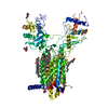 7e2gC  7e2iC C: citing same article ( M: map data used to model this data |
|---|---|
| Similar structure data |


-Links
 PDBj
PDBj
- Assembly
| Deposited unit | 
|
|---|---|
| 1 |
|
-Components
| #1: Protein | Mass: 29497.619 Da / Num. of mol.: 1 Source method: isolated from a genetically manipulated source Source: (gene. exp.) Homo sapiens (human) / Gene: DISP1, DISPAProduction host: Mammalian expression vector EGFP-MCS-pcDNA3.1 (others) References: UniProt: Q96F81 | ||||
|---|---|---|---|---|---|
| #2: Protein | Mass: 140427.594 Da / Num. of mol.: 1 / Mutation: D572N,D573N,D1051N Source method: isolated from a genetically manipulated source Source: (gene. exp.) Homo sapiens (human) / Gene: DISP1, DISPAProduction host: Mammalian expression vector EGFP-MCS-pcDNA3.1 (others) References: UniProt: Q96F81 | ||||
| #3: Chemical | ChemComp-Y01 /   Mass: 486.726 Da / Num. of mol.: 8 / Source method: obtained synthetically / Formula: C31H50O4 Mass: 486.726 Da / Num. of mol.: 8 / Source method: obtained synthetically / Formula: C31H50O4#4: Sugar | ChemComp-NAG / N-Acetylglucosamine  Type: D-saccharide, beta linking / Mass: 221.208 Da / Num. of mol.: 4 / Source method: obtained synthetically / Formula: C8H15NO6 / Feature type: SUBJECT OF INVESTIGATION Type: D-saccharide, beta linking / Mass: 221.208 Da / Num. of mol.: 4 / Source method: obtained synthetically / Formula: C8H15NO6 / Feature type: SUBJECT OF INVESTIGATIONHas ligand of interest | Y | |
-Experimental details
-Experiment
| Experiment | Method: ELECTRON MICROSCOPY |
|---|---|
| EM experiment | Aggregation state: PARTICLE / 3D reconstruction method: single particle reconstruction |
- Sample preparation
| Component | Name: hDisp1NNN-3C / Type: COMPLEX / Entity ID: #1 / Source: RECOMBINANT |
|---|---|
| Molecular weight | Value: 0.15 MDa / Experimental value: NO |
| Source (natural) | Organism: Homo sapiens (human) |
| Source (recombinant) | Organism: Mammalian expression vector EGFP-MCS-pcDNA3.1 (others) |
| Buffer solution | pH: 8 |
| Specimen | Conc.: 7.5 mg/ml / Embedding applied: NO / Shadowing applied: NO / Staining applied: NO / Vitrification applied: YES |
| Vitrification | Cryogen name: ETHANE / Humidity: 100 % |
- Electron microscopy imaging
| Experimental equipment |  Model: Titan Krios / Image courtesy: FEI Company |
|---|---|
| Microscopy | Model: FEI TITAN KRIOS |
| Electron gun | Electron source: FIELD EMISSION GUN / Accelerating voltage: 300 kV / Illumination mode: FLOOD BEAM |
| Electron lens | Mode: BRIGHT FIELDBright-field microscopy |
| Image recording | Electron dose: 50 e/Å2 / Detector mode: SUPER-RESOLUTION / Film or detector model: GATAN K2 SUMMIT (4k x 4k) |
- Processing
| Software | Name: PHENIX / Version: 1.16_3549: / Classification: refinement | ||||||||||||||||||||||||
|---|---|---|---|---|---|---|---|---|---|---|---|---|---|---|---|---|---|---|---|---|---|---|---|---|---|
| CTF correction | Type: PHASE FLIPPING AND AMPLITUDE CORRECTION | ||||||||||||||||||||||||
| 3D reconstruction | Resolution: 3.68 Å / Resolution method: FSC 0.143 CUT-OFF / Num. of particles: 63043 / Symmetry type: POINT | ||||||||||||||||||||||||
| Refine LS restraints |
|
