Movie
Movie Controller

[English] 日本語
 Yorodumi
- PDB-7aoa: Structure of the extended MTA1/HDAC1/MBD2/RBBP4 NURD deacetylase ... -
+ Open data

- Basic information
| Entry | Database: PDB / ID: 7aoa | ||||||||||||
|---|---|---|---|---|---|---|---|---|---|---|---|---|---|
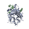
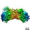
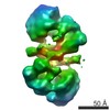
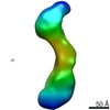
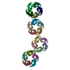
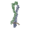
| Title | Structure of the extended MTA1/HDAC1/MBD2/RBBP4 NURD deacetylase complex | ||||||||||||
 Components Components |
| ||||||||||||
 Keywords Keywords |  TRANSCRIPTION / Deacetylase / Complex TRANSCRIPTION / Deacetylase / Complex | ||||||||||||
| Function / homology |  Function and homology information Function and homology informationLoss of MECP2 binding ability to 5mC-DNA / Krueppel-associated box domain binding / Repression of WNT target genes / MECP2 regulates transcription of neuronal ligands / satellite DNA binding / CAF-1 complex / p75NTR negatively regulates cell cycle via SC1 / epidermal cell differentiation / ventricular cardiac muscle tissue development / histone decrotonylase activity ...Loss of MECP2 binding ability to 5mC-DNA / Krueppel-associated box domain binding / Repression of WNT target genes / MECP2 regulates transcription of neuronal ligands / satellite DNA binding / CAF-1 complex / p75NTR negatively regulates cell cycle via SC1 / epidermal cell differentiation / ventricular cardiac muscle tissue development / histone decrotonylase activity / fungiform papilla formation / negative regulation of androgen receptor signaling pathway / NURF complex / regulation of amyloid-beta clearance / regulation of cell fate specification / negative regulation of stem cell population maintenance / DNA replication-dependent chromatin assembly / endoderm development / DNA methylation-dependent heterochromatin formation / NuRD complex / maternal behavior / Transcription of E2F targets under negative control by p107 (RBL1) and p130 (RBL2) in complex with HDAC1 / regulation of stem cell differentiation / ESC/E(Z) complex / protein deacetylation / siRNA binding / Transcription of E2F targets under negative control by DREAM complex / STAT3 nuclear events downstream of ALK signaling / Polo-like kinase mediated events / positive regulation of protein autoubiquitination / histone deacetylase / C2H2 zinc finger domain binding / methyl-CpG binding / protein lysine deacetylase activity / positive regulation of signaling receptor activity / regulation of endopeptidase activity / Hydrolases; Acting on carbon-nitrogen bonds, other than peptide bonds; In linear amides / histone deacetylase activity / embryonic digit morphogenesis / negative regulation of gene expression, epigenetic / positive regulation of oligodendrocyte differentiation / response to ionizing radiation / ATPase complex / positive regulation of stem cell population maintenance / Sin3-type complex / G1/S-Specific Transcription / cellular response to platelet-derived growth factor stimulus / Notch-HLH transcription pathway / eyelid development in camera-type eye / oligodendrocyte differentiation / E-box binding / Transcriptional Regulation by E2F6 / entrainment of circadian clock by photoperiod / locomotor rhythm / odontogenesis of dentin-containing tooth / RNA Polymerase I Transcription Initiation / histone deacetylase complex / SUMOylation of transcription factors / hair follicle placode formation / Regulation of MECP2 expression and activity / cellular response to organic cyclic compound / G0 and Early G1 / positive regulation of Wnt signaling pathway / NF-kappaB binding / negative regulation by host of viral transcription / RNA polymerase II core promoter sequence-specific DNA binding / embryonic organ development / heterochromatin / FOXO-mediated transcription of oxidative stress, metabolic and neuronal genes / negative regulation of intrinsic apoptotic signaling pathway / Cyclin E associated events during G1/S transition / response to mechanical stimulus / negative regulation of canonical NF-kappaB signal transduction / core promoter sequence-specific DNA binding / MECP2 regulates neuronal receptors and channels / Cyclin A:Cdk2-associated events at S phase entry / Deposition of new CENPA-containing nucleosomes at the centromere / Regulation of TP53 Activity through Acetylation / transcription repressor complex / RNA Polymerase I Promoter Opening / SUMOylation of chromatin organization proteins / response to nutrient levels / negative regulation of cell migration / transcription corepressor binding / ERCC6 (CSB) and EHMT2 (G9a) positively regulate rRNA expression / PRC2 methylates histones and DNA / Regulation of PTEN gene transcription / Defective pyroptosis / Deactivation of the beta-catenin transactivating complex / HDACs deacetylate histones / promoter-specific chromatin binding / hippocampus development / Downregulation of SMAD2/3:SMAD4 transcriptional activity / SMAD2/SMAD3:SMAD4 heterotrimer regulates transcription / positive regulation of smooth muscle cell proliferation / negative regulation of transforming growth factor beta receptor signaling pathway / Formation of the beta-catenin:TCF transactivating complex / circadian regulation of gene expression / RUNX1 regulates genes involved in megakaryocyte differentiation and platelet function / brain developmentSimilarity search - Function | ||||||||||||
| Biological species |  Homo sapiens (human) Homo sapiens (human) | ||||||||||||
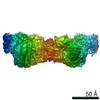
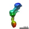
| Method | ELECTRON MICROSCOPY / single particle reconstruction / cryo EM / Resolution: 19.4 Å | ||||||||||||
 Authors Authors | Millard, C.J. / Fairall, L. / Ragan, T.J. / Savva, C.G. / Schwabe, J.W.R. | ||||||||||||
| Funding support |  United Kingdom, 3items United Kingdom, 3items
| ||||||||||||
 Citation Citation | Journal: Nucleic Acids Res / Year: 2020 Title: The topology of chromatin-binding domains in the NuRD deacetylase complex. Authors: Christopher J Millard / Louise Fairall / Timothy J Ragan / Christos G Savva / John W R Schwabe / Abstract: Class I histone deacetylase complexes play essential roles in many nuclear processes. Whilst they contain a common catalytic subunit, they have diverse modes of action determined by associated ...Class I histone deacetylase complexes play essential roles in many nuclear processes. Whilst they contain a common catalytic subunit, they have diverse modes of action determined by associated factors in the distinct complexes. The deacetylase module from the NuRD complex contains three protein domains that control the recruitment of chromatin to the deacetylase enzyme, HDAC1/2. Using biochemical approaches and cryo-electron microscopy, we have determined how three chromatin-binding domains (MTA1-BAH, MBD2/3 and RBBP4/7) are assembled in relation to the core complex so as to facilitate interaction of the complex with the genome. We observe a striking arrangement of the BAH domains suggesting a potential mechanism for binding to di-nucleosomes. We also find that the WD40 domains from RBBP4 are linked to the core with surprising flexibility that is likely important for chromatin engagement. A single MBD2 protein binds asymmetrically to the dimerisation interface of the complex. This symmetry mismatch explains the stoichiometry of the complex. Finally, our structures suggest how the holo-NuRD might assemble on a di-nucleosome substrate. | ||||||||||||
| History |
|
- Structure visualization
| Movie |
Movie viewer |
|---|---|
| Structure viewer | Molecule: MolmilJmol/JSmol |
- Downloads & links
-Download
| PDBx/mmCIF format | 7aoa.cif.gz | 466.6 KB | Display | PDBx/mmCIF format |
|---|---|---|---|---|
| PDB format | pdb7aoa.ent.gz | 365.1 KB | Display | PDB format |
| PDBx/mmJSON format | 7aoa.json.gz | Tree view | PDBx/mmJSON format | |
| Others |  Other downloads Other downloads |
-Validation report
| Arichive directory | https://data.pdbj.org/pub/pdb/validation_reports/ao/7aoaftp://data.pdbj.org/pub/pdb/validation_reports/ao/7aoa | HTTPS FTP |
|---|
-Related structure data
| Related structure data |  11839MC 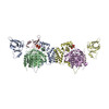 7ao8C 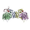 7ao9C M: map data used to model this data C: citing same article ( |
|---|---|
| Similar structure data |

-Links
 PDBj
PDBj


- Assembly
| Deposited unit | 
|
|---|---|
| 1 |
|
-Components
-Protein , 4 types, 7 molecules CDAEBFG
| #1: Protein | / Demethylase / DMTase / Methyl-CpG-binding protein MBD2 Mass: 43323.625 Da / Num. of mol.: 1 Source method: isolated from a genetically manipulated source Source: (gene. exp.) Homo sapiens (human) / Gene: MBD2 / Cell line (production host): HEK293 / Production host: Homo sapiens (human) / References: UniProt: Q9UBB5 | ||||
|---|---|---|---|---|---|
| #2: Protein | Mass: 80904.312 Da / Num. of mol.: 2 Source method: isolated from a genetically manipulated source Source: (gene. exp.) Homo sapiens (human) / Gene: MTA1 / Cell line (production host): HEK293 / Production host: Homo sapiens (human) / References: UniProt: Q13330#3: Protein | HDAC1 / HD1Mass: 55178.906 Da / Num. of mol.: 2 Source method: isolated from a genetically manipulated source Source: (gene. exp.) Homo sapiens (human) / Gene: HDAC1, RPD3L1 / Cell line (production host): HEK293 / Production host: Homo sapiens (human) / References: UniProt: Q13547, histone deacetylase#4: Protein | Mass: 47709.527 Da / Num. of mol.: 2 Source method: isolated from a genetically manipulated source Source: (gene. exp.) Homo sapiens (human) / Gene: RBBP4, RBAP48 / Cell line (production host): HEK293 / Production host: Homo sapiens (human) / References: UniProt: Q09028 |
-Non-polymers , 3 types, 8 molecules 


| #5: Chemical | Phytic acid Mass: 660.035 Da / Num. of mol.: 2 / Source method: obtained synthetically / Formula: C6H18O24P6 Mass: 660.035 Da / Num. of mol.: 2 / Source method: obtained synthetically / Formula: C6H18O24P6#6: Chemical |  Mass: 65.409 Da / Num. of mol.: 2 / Source method: obtained synthetically / Formula: Zn Mass: 65.409 Da / Num. of mol.: 2 / Source method: obtained synthetically / Formula: Zn#7: Chemical | ChemComp-K /  Mass: 39.098 Da / Num. of mol.: 4 / Source method: obtained synthetically / Formula: K Mass: 39.098 Da / Num. of mol.: 4 / Source method: obtained synthetically / Formula: K |
|---|
-Details
| Has ligand of interest | N |
|---|
-Experimental details
-Experiment
| Experiment | Method: ELECTRON MICROSCOPY |
|---|---|
| EM experiment | Aggregation state: PARTICLE / 3D reconstruction method: single particle reconstruction |
- Sample preparation
| Component | Name: Extended NuRD deacetylase complex containing two copies of MTA1, HDAC1 and RBBP4 and a single copy of MBD2 Type: COMPLEX / Entity ID: #1-#4 / Source: RECOMBINANT | ||||||||||||||||||||||||
|---|---|---|---|---|---|---|---|---|---|---|---|---|---|---|---|---|---|---|---|---|---|---|---|---|---|
| Molecular weight | Value: 0.34 MDa / Experimental value: NO | ||||||||||||||||||||||||
| Source (natural) | Organism: Homo sapiens (human) | ||||||||||||||||||||||||
| Source (recombinant) | Organism: Homo sapiens (human) | ||||||||||||||||||||||||
| Buffer solution | pH: 7.5 | ||||||||||||||||||||||||
| Buffer component |
| ||||||||||||||||||||||||
| Specimen | Conc.: 0.1 mg/ml / Embedding applied: NO / Shadowing applied: NO / Staining applied: NO / Vitrification applied: YES | ||||||||||||||||||||||||
| Specimen support | Details: 40 mA for 120 sec / Grid material: GOLD / Grid mesh size: 300 divisions/in. / Grid type: Quantifoil R1.2/1.3 | ||||||||||||||||||||||||
| Vitrification | Instrument: FEI VITROBOT MARK IV / Cryogen name: ETHANE / Humidity: 100 % / Chamber temperature: 277 K / Details: Blot for 3 seconds, blot force 10 |
- Electron microscopy imaging
| Experimental equipment |  Model: Titan Krios / Image courtesy: FEI Company |
|---|---|
| Microscopy | Model: FEI TITAN KRIOS |
| Electron gun | Electron source: FIELD EMISSION GUN / Accelerating voltage: 300 kV / Illumination mode: FLOOD BEAM |
| Electron lens | Mode: BRIGHT FIELDBright-field microscopy / Nominal magnification: 75000 X / Calibrated magnification: 129629 X / Nominal defocus min: 500 nm / Calibrated defocus min: 500 nm / Cs: 2.7 mm / C2 aperture diameter: 50 µm / Alignment procedure: COMA FREE |
| Specimen holder | Cryogen: NITROGEN / Specimen holder model: FEI TITAN KRIOS AUTOGRID HOLDER / Temperature (min): 100 K |
| Image recording | Average exposure time: 60 sec. / Electron dose: 34 e/Å2 / Detector mode: COUNTING / Film or detector model: FEI FALCON III (4k x 4k) / Num. of grids imaged: 1 / Num. of real images: 1902 |
| EM imaging optics | Phase plate: VOLTA PHASE PLATE |
| Image scans | Sampling size: 14 µm / Width: 4096 / Height: 4096 |
- Processing
| EM software |
| ||||||||||||||||||||||||||||||||||||
|---|---|---|---|---|---|---|---|---|---|---|---|---|---|---|---|---|---|---|---|---|---|---|---|---|---|---|---|---|---|---|---|---|---|---|---|---|---|
| CTF correction | Type: PHASE FLIPPING AND AMPLITUDE CORRECTION | ||||||||||||||||||||||||||||||||||||
| Particle selection | Num. of particles selected: 55799 | ||||||||||||||||||||||||||||||||||||
| Symmetry | Point symmetry: C1 (asymmetric) | ||||||||||||||||||||||||||||||||||||
| 3D reconstruction | Resolution: 19.4 Å / Resolution method: FSC 0.143 CUT-OFF / Num. of particles: 10066 / Symmetry type: POINT | ||||||||||||||||||||||||||||||||||||
| Atomic model building | Protocol: RIGID BODY FIT | ||||||||||||||||||||||||||||||||||||
| Atomic model building |
|