 Movie
Movie Controller


+ Open data

- Basic information
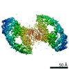
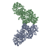
| Entry | Database: PDB / ID: 7aml | ||||||
|---|---|---|---|---|---|---|---|
| Title | RET/GDNF/GFRa1 extracellular complex Cryo-EM structure | ||||||
 Components Components |
| ||||||
 Keywords Keywords |  SIGNALING PROTEIN / VERTEBRATE DEVELOPMENT / PART OF THE RET-GFL-GFRA COMPLEX / NEUROTROPHIC FACTOR SIGNALING PROTEIN / VERTEBRATE DEVELOPMENT / PART OF THE RET-GFL-GFRA COMPLEX / NEUROTROPHIC FACTOR | ||||||
| Function / homology |  Function and homology information Function and homology informationbranchiomeric skeletal muscle development / pronephros morphogenesis / RAF/MAP kinase cascade / : / diencephalon development / positive regulation of ureteric bud formation / postganglionic parasympathetic fiber development / positive regulation of monooxygenase activity / regulation of dopaminergic neuron differentiation / glial cell-derived neurotrophic factor receptor binding ...branchiomeric skeletal muscle development / pronephros morphogenesis / RAF/MAP kinase cascade / : / diencephalon development / positive regulation of ureteric bud formation / postganglionic parasympathetic fiber development / positive regulation of monooxygenase activity / regulation of dopaminergic neuron differentiation / glial cell-derived neurotrophic factor receptor binding / regulation of morphogenesis of a branching structure / regulation of dopamine uptake involved in synaptic transmission / enteric nervous system development / neural crest cell migration involved in autonomic nervous system development / positive regulation of branching involved in ureteric bud morphogenesis / peristalsis / sympathetic nervous system development / peripheral nervous system development / mRNA stabilization / metanephros development / axon extension / positive regulation of kinase activity / neural crest cell migration / branching involved in ureteric bud morphogenesis / homophilic cell adhesion via plasma membrane adhesion molecules / MAP kinase kinase kinase activity / transmembrane receptor protein tyrosine kinase activity / growth factor activity / receptor protein-tyrosine kinase / receptor tyrosine kinase binding / cell surface receptor protein tyrosine kinase signaling pathway / neuron projection development / signaling receptor activity / nervous system development / protein-containing complex assembly / negative regulation of neuron apoptotic process / receptor complex / membrane raft / axon / external side of plasma membrane / calcium ion binding / protein homodimerization activity / positive regulation of transcription by RNA polymerase II / extracellular space / extracellular region / ATP binding / plasma membraneSimilarity search - Function | ||||||
| Biological species |  Danio rerio (zebrafish) Danio rerio (zebrafish) | ||||||
| Method | ELECTRON MICROSCOPY / single particle reconstruction / cryo EM / Resolution: 3.5 Å | ||||||
 Authors Authors | Adams, S.E. / Earl, C.P. / Purkiss, A.G. / McDonald, N.Q. | ||||||
| Funding support |  United Kingdom, 1items United Kingdom, 1items
| ||||||
 Citation Citation | Journal: Structure / Year: 2021 Title: A two-site flexible clamp mechanism for RET-GDNF-GFRα1 assembly reveals both conformational adaptation and strict geometric spacing. Authors: Sarah E Adams / Andrew G Purkiss / Phillip P Knowles / Andrea Nans / David C Briggs / Annabel Borg / Christopher P Earl / Kerry M Goodman / Agata Nawrotek / Aaron J Borg / Pauline B McIntosh ...Authors: Sarah E Adams / Andrew G Purkiss / Phillip P Knowles / Andrea Nans / David C Briggs / Annabel Borg / Christopher P Earl / Kerry M Goodman / Agata Nawrotek / Aaron J Borg / Pauline B McIntosh / Francesca M Houghton / Svend Kjær / Neil Q McDonald / Abstract: RET receptor tyrosine kinase plays vital developmental and neuroprotective roles in metazoans. GDNF family ligands (GFLs) when bound to cognate GFRα co-receptors recognize and activate RET ...RET receptor tyrosine kinase plays vital developmental and neuroprotective roles in metazoans. GDNF family ligands (GFLs) when bound to cognate GFRα co-receptors recognize and activate RET stimulating its cytoplasmic kinase function. The principles for RET ligand-co-receptor recognition are incompletely understood. Here, we report a crystal structure of the cadherin-like module (CLD1-4) from zebrafish RET revealing interdomain flexibility between CLD2 and CLD3. Comparison with a cryo-electron microscopy structure of a ligand-engaged zebrafish RET-GDNF-GFRα1a complex indicates conformational changes within a clade-specific CLD3 loop adjacent to the co-receptor. Our observations indicate that RET is a molecular clamp with a flexible calcium-dependent arm that adapts to different GFRα co-receptors, while its rigid arm recognizes a GFL dimer to align both membrane-proximal cysteine-rich domains. We also visualize linear arrays of RET-GDNF-GFRα1a suggesting that a conserved contact stabilizes higher-order species. Our study reveals that ligand-co-receptor recognition by RET involves both receptor plasticity and strict spacing of receptor dimers by GFL ligands. | ||||||
| History |
|
- Structure visualization
| Movie |
Movie viewer |
|---|---|
| Structure viewer | Molecule: MolmilJmol/JSmol |
- Downloads & links
-Download
| PDBx/mmCIF format | 7aml.cif.gz | 652.5 KB | Display | PDBx/mmCIF format |
|---|---|---|---|---|
| PDB format | pdb7aml.ent.gz | 562 KB | Display | PDB format |
| PDBx/mmJSON format | 7aml.json.gz | Tree view | PDBx/mmJSON format | |
| Others |  Other downloads Other downloads |
-Validation report
| Arichive directory | https://data.pdbj.org/pub/pdb/validation_reports/am/7amlftp://data.pdbj.org/pub/pdb/validation_reports/am/7aml | HTTPS FTP |
|---|
-Related structure data
| Related structure data | 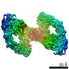 11822MC  7ab8C  7amkC C: citing same article ( M: map data used to model this data |
|---|---|
| Similar structure data |


-Links
 PDBj
PDBj


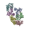
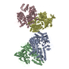
- Assembly
| Deposited unit | 
| |||||||||||||||||||||||||||||||||||||||||||||||||||||||||||||||||||||||||||||||||||||||||||||||||||||||||||||||||||||||||||
|---|---|---|---|---|---|---|---|---|---|---|---|---|---|---|---|---|---|---|---|---|---|---|---|---|---|---|---|---|---|---|---|---|---|---|---|---|---|---|---|---|---|---|---|---|---|---|---|---|---|---|---|---|---|---|---|---|---|---|---|---|---|---|---|---|---|---|---|---|---|---|---|---|---|---|---|---|---|---|---|---|---|---|---|---|---|---|---|---|---|---|---|---|---|---|---|---|---|---|---|---|---|---|---|---|---|---|---|---|---|---|---|---|---|---|---|---|---|---|---|---|---|---|---|---|
| 1 |
| |||||||||||||||||||||||||||||||||||||||||||||||||||||||||||||||||||||||||||||||||||||||||||||||||||||||||||||||||||||||||||
| Noncrystallographic symmetry (NCS) | NCS domain:
NCS domain segments:
|