 Movie
Movie Controller

[English] 日本語
 Yorodumi
- PDB-7aeb: Cryo-EM structure of an extracellular contractile injection syste... -
+ Open data

- Basic information
| Entry | Database: PDB / ID: 7aeb | ||||||||||||
|---|---|---|---|---|---|---|---|---|---|---|---|---|---|
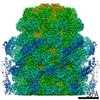
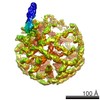
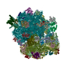
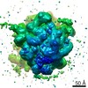
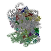
| Title | Cryo-EM structure of an extracellular contractile injection system in marine bacterium Algoriphagus machipongonensis, the baseplate complex in extended state applied 6-fold symmetry. | ||||||||||||
 Components Components |
| ||||||||||||
 Keywords Keywords |  STRUCTURAL PROTEIN / extracellular contractile injection system STRUCTURAL PROTEIN / extracellular contractile injection system | ||||||||||||
| Function / homology |  Function and homology information Function and homology information | ||||||||||||
| Biological species |  Algoriphagus machipongonensis (bacteria) Algoriphagus machipongonensis (bacteria) | ||||||||||||
| Method | ELECTRON MICROSCOPY / single particle reconstruction / cryo EM / Resolution: 2.7 Å | ||||||||||||
 Authors Authors | Xu, J. / Ericson, C. / Feldmueller, M. / Lien, Y.W. / Pilhofer, M. | ||||||||||||
| Funding support |  Switzerland, 3items Switzerland, 3items
| ||||||||||||
 Citation Citation | Journal: Nat Microbiol / Year: 2022 Title: Identification and structure of an extracellular contractile injection system from the marine bacterium Algoriphagus machipongonensis. Authors: Jingwei Xu / Charles F Ericson / Yun-Wei Lien / Florentine U N Rutaganira / Fabian Eisenstein / Miki Feldmüller / Nicole King / Martin Pilhofer /   Abstract: Contractile injection systems (CISs) are phage tail-like nanomachines, mediating bacterial cell-cell interactions as either type VI secretion systems (T6SSs) or extracellular CISs (eCISs). ...Contractile injection systems (CISs) are phage tail-like nanomachines, mediating bacterial cell-cell interactions as either type VI secretion systems (T6SSs) or extracellular CISs (eCISs). Bioinformatic studies uncovered a phylogenetic group of hundreds of putative CIS gene clusters that are highly diverse and widespread; however, only four systems have been characterized. Here we studied a putative CIS gene cluster in the marine bacterium Algoriphagus machipongonensis. Using an integrative approach, we show that the system is compatible with an eCIS mode of action. Our cryo-electron microscopy structure revealed several features that differ from those seen in other CISs: a 'cap adaptor' located at the distal end, a 'plug' exposed to the tube lumen, and a 'cage' formed by massive extensions of the baseplate. These elements are conserved in other CISs, and our genetic tools identified that they are required for assembly, cargo loading and function. Furthermore, our atomic model highlights specific evolutionary hotspots and will serve as a framework for understanding and re-engineering CISs. | ||||||||||||
| History |
|
- Structure visualization
| Movie |
Movie viewer |
|---|---|
| Structure viewer | Molecule: MolmilJmol/JSmol |
- Downloads & links
-Download
| PDBx/mmCIF format | 7aeb.cif.gz | 2.8 MB | Display | PDBx/mmCIF format |
|---|---|---|---|---|
| PDB format | pdb7aeb.ent.gz | Display | PDB format | |
| PDBx/mmJSON format | 7aeb.json.gz | Tree view | PDBx/mmJSON format | |
| Others |  Other downloads Other downloads |
-Validation report
| Arichive directory | https://data.pdbj.org/pub/pdb/validation_reports/ae/7aebftp://data.pdbj.org/pub/pdb/validation_reports/ae/7aeb | HTTPS FTP |
|---|
-Related structure data
| Related structure data |  11743MC 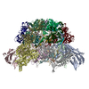 7adzC  7ae0C 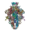 7aefC  7aekC C: citing same article ( M: map data used to model this data |
|---|---|
| Similar structure data |


-Links
 PDBj
PDBj
- Assembly
| Deposited unit | 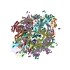
|
|---|---|
| 1 |
|
-Components
-Protein , 7 types, 42 molecules ABCDEFGHIJKLMNOPQRSTUVWXYZabcd...
| #1: Protein | Tripod (photography) / baseplate protein (Algo12) Mass: 107770.953 Da / Num. of mol.: 6 / Source method: isolated from a natural source Details: the residues (552-end) could not be built up due to the poor map density. Source: (natural) Algoriphagus machipongonensis (bacteria) / References: UniProt: A3HTB3#2: Protein | Mass: 119379.320 Da / Num. of mol.: 6 / Source method: isolated from a natural source Details: The C terminal part (residues 916-1012aa) is built up by poly-alanine chain due to the poor map density. Source: (natural) Algoriphagus machipongonensis (bacteria) / References: UniProt: A3HTB4#3: Protein | Mass: 26571.104 Da / Num. of mol.: 6 / Source method: isolated from a natural source Details: The residue S169 is not found in relative map, which might be cleaved in the structure. Source: (natural) Algoriphagus machipongonensis (bacteria) / References: UniProt: A3HTB8#4: Protein | Mass: 15762.021 Da / Num. of mol.: 6 / Source method: isolated from a natural source / Source: (natural) Algoriphagus machipongonensis (bacteria) / References: UniProt: A3HTB5#5: Protein | Mass: 17063.238 Da / Num. of mol.: 6 / Source method: isolated from a natural source / Source: (natural) Algoriphagus machipongonensis (bacteria) / References: UniProt: A3HTC0#6: Protein | Mass: 76319.039 Da / Num. of mol.: 6 / Source method: isolated from a natural source Details: the residues 288-320aa could not be built up in the model, with residues 430-446aa assigned with poly-alanine chain. Source: (natural) Algoriphagus machipongonensis (bacteria) / References: UniProt: A3HTC2#7: Protein | Mass: 16375.458 Da / Num. of mol.: 6 / Source method: isolated from a natural source / Source: (natural) Algoriphagus machipongonensis (bacteria) / References: UniProt: A3HTC1 |
|---|
-Experimental details
-Experiment
| Experiment | Method: ELECTRON MICROSCOPY |
|---|---|
| EM experiment | Aggregation state: PARTICLE / 3D reconstruction method: single particle reconstruction |
- Sample preparation
| Component | Name: The baseplate complex of an extracellular contractile injection system in marine bacterium Algoriphagus machipongonensis, applied 6-fold symmetry. Type: COMPLEX / Entity ID: all / Source: NATURAL |
|---|---|
| Source (natural) | Organism: Algoriphagus machipongonensis (bacteria) |
| Buffer solution | pH: 8 |
| Specimen | Embedding applied: NO / Shadowing applied: NO / Staining applied: NO / Vitrification applied: YES |
| Vitrification | Instrument: FEI VITROBOT MARK IV / Cryogen name: ETHANE-PROPANE |
- Electron microscopy imaging
| Experimental equipment |  Model: Titan Krios / Image courtesy: FEI Company |
|---|---|
| Microscopy | Model: FEI TITAN KRIOS |
| Electron gun | Electron source: FIELD EMISSION GUN / Accelerating voltage: 300 kV / Illumination mode: FLOOD BEAM |
| Electron lens | Mode: BRIGHT FIELDBright-field microscopy |
| Image recording | Electron dose: 60 e/Å2 / Film or detector model: GATAN K3 (6k x 4k) |
- Processing
| CTF correction | Type: PHASE FLIPPING AND AMPLITUDE CORRECTION |
|---|---|
| 3D reconstruction | Resolution: 2.7 Å / Resolution method: FSC 0.143 CUT-OFF / Num. of particles: 82969 / Symmetry type: POINT |
| Atomic model building | Protocol: AB INITIO MODEL / Space: REAL |
