Movie
Movie Controller


+ Open data

- Basic information
| Entry | Database: PDB / ID: 6zqi | ||||||||||||||||||
|---|---|---|---|---|---|---|---|---|---|---|---|---|---|---|---|---|---|---|---|
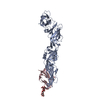
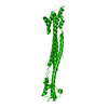
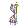
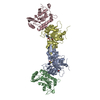
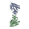
| Title | Cryo-EM structure of Spondweni virus prME | ||||||||||||||||||
 Components Components | (Genome polyprotein) x 2 | ||||||||||||||||||
 Keywords Keywords |  VIRUS / Flavivirus VIRUS / Flavivirus | ||||||||||||||||||
| Function / homology |  Function and homology information Function and homology informationsymbiont-mediated suppression of host JAK-STAT cascade via inhibition of STAT2 activity / ribonucleoside triphosphate phosphatase activity / viral capsid / double-stranded RNA binding / mRNA (nucleoside-2'-O-)-methyltransferase activity / mRNA 5'-cap (guanine-N7-)-methyltransferase activity / RNA helicase activity / host cell endoplasmic reticulum membrane / protein dimerization activity / symbiont entry into host cell ...symbiont-mediated suppression of host JAK-STAT cascade via inhibition of STAT2 activity / ribonucleoside triphosphate phosphatase activity / viral capsid / double-stranded RNA binding / mRNA (nucleoside-2'-O-)-methyltransferase activity / mRNA 5'-cap (guanine-N7-)-methyltransferase activity / RNA helicase activity / host cell endoplasmic reticulum membrane / protein dimerization activity / symbiont entry into host cell / viral RNA genome replication / RNA-dependent RNA polymerase activity / serine-type endopeptidase activity / fusion of virus membrane with host endosome membrane / symbiont-mediated suppression of host type I interferon-mediated signaling pathway / host cell nucleus / virion attachment to host cell / virion membrane / structural molecule activity / proteolysis / extracellular region / ATP binding / membrane / metal ion bindingSimilarity search - Function | ||||||||||||||||||
| Biological species |  Spondweni virus Spondweni virus | ||||||||||||||||||
| Method | ELECTRON MICROSCOPY / single particle reconstruction / cryo EM / Resolution: 3.8 Å | ||||||||||||||||||
 Authors Authors | Renner, M. / Dejnirattisai, W. / Carrique, L. / Serna Martin, I. / Karia, D. / Ilca, S.L. / Ho, S.F. / Kotecha, A. / Keown, J.R. / Mongkolsapaya, J. ...Renner, M. / Dejnirattisai, W. / Carrique, L. / Serna Martin, I. / Karia, D. / Ilca, S.L. / Ho, S.F. / Kotecha, A. / Keown, J.R. / Mongkolsapaya, J. / Screaton, G.R. / Grimes, J.M. | ||||||||||||||||||
| Funding support |  United Kingdom, 5items United Kingdom, 5items
| ||||||||||||||||||
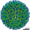
 Citation Citation | Journal: Nat Commun / Year: 2021 Title: Flavivirus maturation leads to the formation of an occupied lipid pocket in the surface glycoproteins. Authors: Max Renner / Wanwisa Dejnirattisai / Loïc Carrique / Itziar Serna Martin / Dimple Karia / Serban L Ilca / Shu F Ho / Abhay Kotecha / Jeremy R Keown / Juthathip Mongkolsapaya / Gavin R ...Authors: Max Renner / Wanwisa Dejnirattisai / Loïc Carrique / Itziar Serna Martin / Dimple Karia / Serban L Ilca / Shu F Ho / Abhay Kotecha / Jeremy R Keown / Juthathip Mongkolsapaya / Gavin R Screaton / Jonathan M Grimes /   Abstract: Flaviviruses such as Dengue (DENV) or Zika virus (ZIKV) assemble into an immature form within the endoplasmatic reticulum (ER), and are then processed by furin protease in the trans-Golgi. To better ...Flaviviruses such as Dengue (DENV) or Zika virus (ZIKV) assemble into an immature form within the endoplasmatic reticulum (ER), and are then processed by furin protease in the trans-Golgi. To better grasp maturation, we carry out cryo-EM reconstructions of immature Spondweni virus (SPOV), a human flavivirus of the same serogroup as ZIKV. By employing asymmetric localised reconstruction we push the resolution to 3.8 Å, enabling us to refine an atomic model which includes the crucial furin protease recognition site and a conserved Histidine pH-sensor. For direct comparison, we also solve structures of the mature forms of SPONV and DENV to 2.6 Å and 3.1 Å, respectively. We identify an ordered lipid that is present in only the mature forms of ZIKV, SPOV, and DENV and can bind as a consequence of rearranging amphipathic stem-helices of E during maturation. We propose a structural role for the pocket and suggest it stabilizes mature E. | ||||||||||||||||||
| History |
|
- Structure visualization
| Movie |
Movie viewer |
|---|---|
| Structure viewer | Molecule: MolmilJmol/JSmol |
- Downloads & links
-Download
| PDBx/mmCIF format | 6zqi.cif.gz | 122.8 KB | Display | PDBx/mmCIF format |
|---|---|---|---|---|
| PDB format | pdb6zqi.ent.gz | 98 KB | Display | PDB format |
| PDBx/mmJSON format | 6zqi.json.gz | Tree view | PDBx/mmJSON format | |
| Others |  Other downloads Other downloads |
-Validation report
| Arichive directory | https://data.pdbj.org/pub/pdb/validation_reports/zq/6zqiftp://data.pdbj.org/pub/pdb/validation_reports/zq/6zqi | HTTPS FTP |
|---|
-Related structure data
| Related structure data |  11364MC  6zqjC  6zquC  6zqvC  6zqwC M: map data used to model this data C: citing same article ( |
|---|---|
| Similar structure data |


-Links
 PDBj
PDBj

- Assembly
| Deposited unit | 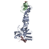
|
|---|---|
| 1 |
|
-Components
| #1: Protein | Mass: 54838.207 Da / Num. of mol.: 1 / Source method: isolated from a natural source / Source: (natural) Spondweni virus / References: UniProt: C8XPB6 | ||||
|---|---|---|---|---|---|
| #2: Protein | Mass: 19070.752 Da / Num. of mol.: 2 / Source method: isolated from a natural source / Source: (natural) Spondweni virus / References: UniProt: C8XPB6#3: Sugar | ChemComp-NAG / | N-Acetylglucosamine  Type: D-saccharide, beta linking / Mass: 221.208 Da / Num. of mol.: 1 Type: D-saccharide, beta linking / Mass: 221.208 Da / Num. of mol.: 1Source method: isolated from a genetically manipulated source Formula: C8H15NO6 Has ligand of interest | N | |
-Experimental details
-Experiment
| Experiment | Method: ELECTRON MICROSCOPY |
|---|---|
| EM experiment | Aggregation state: PARTICLE / 3D reconstruction method: single particle reconstruction |
- Sample preparation
| Component | Name: Spondweni virus / Type: VIRUS / Details: Virus cultivated in C6/36 cells / Entity ID: #1-#2 / Source: NATURAL |
|---|---|
| Molecular weight | Experimental value: NO |
| Source (natural) | Organism: Spondweni virus |
| Details of virus | Empty: NO / Enveloped: YES / Isolate: OTHER / Type: VIRION |
| Natural host | Organism: Aedes circumluteolus |
| Buffer solution | pH: 7.4 / Details: PBS buffer |
| Specimen | Embedding applied: NO / Shadowing applied: NO / Staining applied: NO / Vitrification applied: YES / Details: UV inactivated |
| Specimen support | Grid material: GOLD / Grid mesh size: 200 divisions/in. / Grid type: Quantifoil R2/1 |
| Vitrification | Instrument: FEI VITROBOT MARK IV / Cryogen name: ETHANE-PROPANE |
- Electron microscopy imaging
| Experimental equipment |  Model: Titan Krios / Image courtesy: FEI Company |
|---|---|
| Microscopy | Model: FEI TITAN KRIOS |
| Electron gun | Electron source: FIELD EMISSION GUN / Accelerating voltage: 300 kV / Illumination mode: FLOOD BEAM |
| Electron lens | Mode: BRIGHT FIELDBright-field microscopy / Cs: 2.7 mm |
| Image recording | Electron dose: 30 e/Å2 / Film or detector model: GATAN K3 (6k x 4k) |
- Processing
| Software | Name: PHENIX / Version: 1.17.1_3660: / Classification: refinement | ||||||||||||||||||||||||
|---|---|---|---|---|---|---|---|---|---|---|---|---|---|---|---|---|---|---|---|---|---|---|---|---|---|
| EM software |
| ||||||||||||||||||||||||
| CTF correction | Type: PHASE FLIPPING AND AMPLITUDE CORRECTION | ||||||||||||||||||||||||
| Particle selection | Num. of particles selected: 2572800 Details: 2572800 sub-particles were generated from 42880 immature Spondweni virions (selected in cryoSPARC) via localised reconstruction | ||||||||||||||||||||||||
| Symmetry | Point symmetry: C1 (asymmetric) | ||||||||||||||||||||||||
| 3D reconstruction | Resolution: 3.8 Å / Resolution method: FSC 0.143 CUT-OFF / Num. of particles: 281619 / Symmetry type: POINT | ||||||||||||||||||||||||
| Atomic model building | Space: REAL | ||||||||||||||||||||||||
| Atomic model building | PDB-ID: 3C5X | ||||||||||||||||||||||||
| Refine LS restraints |
|