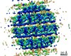
Movie
Movie Controller

[English] 日本語
 Yorodumi
- PDB-6z5l: Helical reconstruction of influenza A virus M1 in complex with nu... -
+ Open data

- Basic information
| Entry | Database: PDB / ID: 6z5l | ||||||||||||
|---|---|---|---|---|---|---|---|---|---|---|---|---|---|
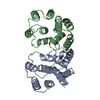
| Title | Helical reconstruction of influenza A virus M1 in complex with nucleic acid. | ||||||||||||
 Components Components | Matrix protein 1 | ||||||||||||
 Keywords Keywords |  VIRAL PROTEIN / M1 / Matrix protein / Influenza virus / Assembly / ribonucleoprotein complex VIRAL PROTEIN / M1 / Matrix protein / Influenza virus / Assembly / ribonucleoprotein complex | ||||||||||||
| Function / homology |  Function and homology information Function and homology informationAssembly of Viral Components at the Budding Site / Influenza Infection / Fusion of the Influenza Virion to the Host Cell Endosome / Release / Budding / Packaging of Eight RNA Segments / Uncoating of the Influenza Virion / Entry of Influenza Virion into Host Cell via Endocytosis / Viral RNP Complexes in the Host Cell Nucleus / NEP/NS2 Interacts with the Cellular Export Machinery ...Assembly of Viral Components at the Budding Site / Influenza Infection / Fusion of the Influenza Virion to the Host Cell Endosome / Release / Budding / Packaging of Eight RNA Segments / Uncoating of the Influenza Virion / Entry of Influenza Virion into Host Cell via Endocytosis / Viral RNP Complexes in the Host Cell Nucleus / NEP/NS2 Interacts with the Cellular Export Machinery / Viral mRNA Translation / viral budding from plasma membrane / structural constituent of virion / host cell nucleus / virion membrane / RNA binding / extracellular region / plasma membraneSimilarity search - Function | ||||||||||||
| Biological species |   Influenza A virus Influenza A virus | ||||||||||||
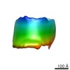
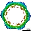
| Method | ELECTRON MICROSCOPY / helical reconstruction / cryo EM / Resolution: 3.8 Å | ||||||||||||
 Authors Authors | Xiong, X. / Qu, K. / Briggs, J.A.G. | ||||||||||||
| Funding support |  United Kingdom, European Union, United Kingdom, European Union,  Germany, 3items Germany, 3items
| ||||||||||||
 Citation Citation | Journal: Nature / Year: 2020 Title: The native structure of the assembled matrix protein 1 of influenza A virus. Authors: Julia Peukes / Xiaoli Xiong / Simon Erlendsson / Kun Qu / William Wan / Leslie J Calder / Oliver Schraidt / Susann Kummer / Stefan M V Freund / Hans-Georg Kräusslich / John A G Briggs /    Abstract: Influenza A virus causes millions of severe cases of disease during annual epidemics. The most abundant protein in influenza virions is matrix protein 1 (M1), which mediates virus assembly by ...Influenza A virus causes millions of severe cases of disease during annual epidemics. The most abundant protein in influenza virions is matrix protein 1 (M1), which mediates virus assembly by forming an endoskeleton beneath the virus membrane. The structure of full-length M1, and how it oligomerizes to mediate the assembly of virions, is unknown. Here we determine the complete structure of assembled M1 within intact virus particles, as well as the structure of M1 oligomers reconstituted in vitro. We find that the C-terminal domain of M1 is disordered in solution but can fold and bind in trans to the N-terminal domain of another M1 monomer, thus polymerizing M1 into linear strands that coat the interior surface of the membrane of the assembling virion. In the M1 polymer, five histidine residues-contributed by three different monomers of M1-form a cluster that can serve as the pH-sensitive disassembly switch after entry into a target cell. These structures therefore reveal mechanisms of influenza virus assembly and disassembly. #1: Journal: Biorxiv / Year: 2020Title: The native structure of the full-length, assembled influenza A virus matrix protein, M1. Authors: Peukes, J. / Xiong, X. / Erlendsson, S. / Qu, K. / Wan, W. / Calder, L.J. / Schraidt, O. / Kummer, S. / Freund, S.M.V. / Krausslich, H.G. / Briggs, J.A.G. | ||||||||||||
| History |
|
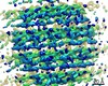
- Structure visualization
| Movie |
Movie viewer |
|---|---|
| Structure viewer | Molecule: MolmilJmol/JSmol |
- Downloads & links
-Download
| PDBx/mmCIF format | 6z5l.cif.gz | 55.9 KB | Display | PDBx/mmCIF format |
|---|---|---|---|---|
| PDB format | pdb6z5l.ent.gz | 43.2 KB | Display | PDB format |
| PDBx/mmJSON format | 6z5l.json.gz | Tree view | PDBx/mmJSON format | |
| Others |  Other downloads Other downloads |
-Validation report
| Arichive directory | https://data.pdbj.org/pub/pdb/validation_reports/z5/6z5lftp://data.pdbj.org/pub/pdb/validation_reports/z5/6z5l | HTTPS FTP |
|---|
-Related structure data
| Related structure data | 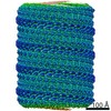 11079MC  6z5jC M: map data used to model this data C: citing same article ( |
|---|---|
| Similar structure data |
-Links
 PDBj
PDBj


- Assembly
| Deposited unit | 
|
|---|---|
| 1 | x 80
|
-Components
| #1: Protein | Mass: 28971.430 Da / Num. of mol.: 1 / Mutation: R134K Source method: isolated from a genetically manipulated source Source: (gene. exp.) Influenza A virus (strain A/Puerto Rico/8/1934 H1N1)Variant: R134K / Plasmid: pET21b / Production host:  Escherichia coli BL21(DE3) (bacteria) / Variant (production host): Rosetta2 / References: UniProt: P03485 Escherichia coli BL21(DE3) (bacteria) / Variant (production host): Rosetta2 / References: UniProt: P03485 |
|---|
-Experimental details
-Experiment
| Experiment | Method: ELECTRON MICROSCOPY |
|---|---|
| EM experiment | Aggregation state: HELICAL ARRAY / 3D reconstruction method: helical reconstruction |
- Sample preparation
| Component | Name: Helical reconstruction of influenza A virus M1 protein in complex with nucleic acid Type: COMPLEX / Details: Influenza A M1 in complex with 6.4 Kb DNA plasmid / Entity ID: all / Source: RECOMBINANT | ||||||||||||||||
|---|---|---|---|---|---|---|---|---|---|---|---|---|---|---|---|---|---|
| Molecular weight |
| ||||||||||||||||
| Source (natural) | Organism: Influenza A virus (strain A/Puerto Rico/8/1934 H1N1) | ||||||||||||||||
| Source (recombinant) | Organism: Escherichia coli BL21(DE3) (bacteria) / Strain: Rosetta 2 / Plasmid: pET21b | ||||||||||||||||
| Buffer solution | pH: 10 | ||||||||||||||||
| Buffer component |
| ||||||||||||||||
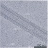
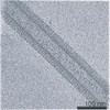
| Specimen | Conc.: 0.1 mg/ml / Embedding applied: NO / Shadowing applied: NO / Staining applied: NO / Vitrification applied: YES / Details: M1 at 0.1 mg/ml plasmid DNA at 0.25 mg/ml | ||||||||||||||||
| Specimen support | Grid material: COPPER / Grid mesh size: 200 divisions/in. / Grid type: C-flat-2/2 | ||||||||||||||||
| Vitrification | Instrument: FEI VITROBOT MARK IV / Cryogen name: ETHANE / Humidity: 100 % / Chamber temperature: 298 K Details: sample was applied 3 times each with 30s adsorption time |
- Electron microscopy imaging
| Experimental equipment |  Model: Titan Krios / Image courtesy: FEI Company |
|---|---|
| Microscopy | Model: FEI TITAN KRIOS |
| Electron gun | Electron source: FIELD EMISSION GUN / Accelerating voltage: 300 kV / Illumination mode: FLOOD BEAM |
| Electron lens | Mode: BRIGHT FIELDBright-field microscopy / Nominal magnification: 105000 X / Nominal defocus max: 3000 nm / Nominal defocus min: 1000 nm / Cs: 2.7 mm / Alignment procedure: COMA FREE |
| Specimen holder | Cryogen: NITROGEN / Specimen holder model: FEI TITAN KRIOS AUTOGRID HOLDER |
| Image recording | Electron dose: 47.1 e/Å2 / Detector mode: COUNTING / Film or detector model: GATAN K2 SUMMIT (4k x 4k) / Num. of grids imaged: 1 / Num. of real images: 2347 |
| EM imaging optics | Energyfilter name: GIF Quantum LS / Energyfilter slit width: 20 eV |
- Processing
| Software | Name: PHENIX / Version: 1.17.1_3660: / Classification: refinement | ||||||||||||||||||||||||||||||||||||||||||||||||||
|---|---|---|---|---|---|---|---|---|---|---|---|---|---|---|---|---|---|---|---|---|---|---|---|---|---|---|---|---|---|---|---|---|---|---|---|---|---|---|---|---|---|---|---|---|---|---|---|---|---|---|---|
| EM software |
| ||||||||||||||||||||||||||||||||||||||||||||||||||
| CTF correction | Type: PHASE FLIPPING AND AMPLITUDE CORRECTION | ||||||||||||||||||||||||||||||||||||||||||||||||||
| Helical symmerty | Angular rotation/subunit: -11.1081 ° / Axial rise/subunit: 3.08413 Å / Axial symmetry: D1 | ||||||||||||||||||||||||||||||||||||||||||||||||||
| Particle selection | Num. of particles selected: 463152 / Details: Manual picking | ||||||||||||||||||||||||||||||||||||||||||||||||||
| 3D reconstruction | Resolution: 3.8 Å / Resolution method: FSC 0.143 CUT-OFF / Num. of particles: 17984 / Algorithm: FOURIER SPACE / Num. of class averages: 1 / Symmetry type: HELICAL | ||||||||||||||||||||||||||||||||||||||||||||||||||
| Atomic model building | B value: 37.9 / Protocol: AB INITIO MODEL / Space: REAL / Target criteria: correlation coefficient | ||||||||||||||||||||||||||||||||||||||||||||||||||
| Atomic model building | PDB-ID: 1AA7 Pdb chain-ID: A | ||||||||||||||||||||||||||||||||||||||||||||||||||
| Refine LS restraints |
|