 Movie
Movie Controller

[English] 日本語
 Yorodumi
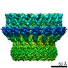
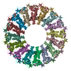
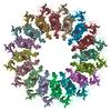
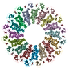
- PDB-6sb3: CryoEM structure of murine perforin-2 ectodomain in a pre-pore form -
+ Open data

- Basic information
| Entry | Database: PDB / ID: 6sb3 | ||||||
|---|---|---|---|---|---|---|---|
| Title | CryoEM structure of murine perforin-2 ectodomain in a pre-pore form | ||||||
 Components Components | Macrophage-expressed gene 1 protein | ||||||
 Keywords Keywords |  TOXIN / pore-forming protein / pre-pore / MACPF TOXIN / pore-forming protein / pre-pore / MACPF | ||||||
| Function / homology |  Function and homology information Function and homology informationdendritic cell antigen processing and presentation / antigen processing and presentation of exogenous peptide antigen / phagolysosome membrane / endolysosome / pore-forming activity / antibacterial innate immune response / wide pore channel activity / antigen processing and presentation of exogenous peptide antigen via MHC class I / phagocytic vesicle / phagocytic vesicle membrane ...dendritic cell antigen processing and presentation / antigen processing and presentation of exogenous peptide antigen / phagolysosome membrane / endolysosome / pore-forming activity / antibacterial innate immune response / wide pore channel activity / antigen processing and presentation of exogenous peptide antigen via MHC class I / phagocytic vesicle / phagocytic vesicle membrane / cytoplasmic vesicle / defense response to Gram-negative bacterium / adaptive immune response / defense response to Gram-positive bacterium / defense response to bacteriumSimilarity search - Function | ||||||
| Biological species |  Mus musculus (house mouse) Mus musculus (house mouse) | ||||||
| Method | ELECTRON MICROSCOPY / single particle reconstruction / cryo EM / Resolution: 3.5 Å | ||||||
 Authors Authors | Ni, T. / Yu, X. / Gilbert, R.J.C. | ||||||
| Funding support |  United Kingdom, 1items United Kingdom, 1items
| ||||||
 Citation Citation | Journal: Sci Adv / Year: 2020 Title: Structure and mechanism of bactericidal mammalian perforin-2, an ancient agent of innate immunity. Authors: Tao Ni / Fang Jiao / Xiulian Yu / Saša Aden / Lucy Ginger / Sophie I Williams / Fangfang Bai / Vojtěch Pražák / Dimple Karia / Phillip Stansfeld / Peijun Zhang / George Munson / Gregor ...Authors: Tao Ni / Fang Jiao / Xiulian Yu / Saša Aden / Lucy Ginger / Sophie I Williams / Fangfang Bai / Vojtěch Pražák / Dimple Karia / Phillip Stansfeld / Peijun Zhang / George Munson / Gregor Anderluh / Simon Scheuring / Robert J C Gilbert /   Abstract: Perforin-2 (MPEG1) is thought to enable the killing of invading microbes engulfed by macrophages and other phagocytes, forming pores in their membranes. Loss of perforin-2 renders individual ...Perforin-2 (MPEG1) is thought to enable the killing of invading microbes engulfed by macrophages and other phagocytes, forming pores in their membranes. Loss of perforin-2 renders individual phagocytes and whole organisms significantly more susceptible to bacterial pathogens. Here, we reveal the mechanism of perforin-2 activation and activity using atomic structures of pre-pore and pore assemblies, high-speed atomic force microscopy, and functional assays. Perforin-2 forms a pre-pore assembly in which its pore-forming domain points in the opposite direction to its membrane-targeting domain. Acidification then triggers pore formation, via a 180° conformational change. This novel and unexpected mechanism prevents premature bactericidal attack and may have played a key role in the evolution of all perforin family proteins. | ||||||
| History |
|
- Structure visualization
| Movie |
Movie viewer |
|---|---|
| Structure viewer | Molecule: MolmilJmol/JSmol |
- Downloads & links
-Download
| PDBx/mmCIF format | 6sb3.cif.gz | 1.5 MB | Display | PDBx/mmCIF format |
|---|---|---|---|---|
| PDB format | pdb6sb3.ent.gz | 1.3 MB | Display | PDB format |
| PDBx/mmJSON format | 6sb3.json.gz | Tree view | PDBx/mmJSON format | |
| Others |  Other downloads Other downloads |
-Validation report
| Arichive directory | https://data.pdbj.org/pub/pdb/validation_reports/sb/6sb3ftp://data.pdbj.org/pub/pdb/validation_reports/sb/6sb3 | HTTPS FTP |
|---|
-Related structure data
| Related structure data | 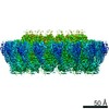 10134MC  6sb1C  6sb4C  6sb5C M: map data used to model this data C: citing same article ( |
|---|---|
| Similar structure data |


-Links
 PDBj
PDBj- Assembly
| Deposited unit | 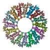
|
|---|---|
| 1 |
|
-Components
| #1: Protein | Mass: 71129.531 Da / Num. of mol.: 16 Source method: isolated from a genetically manipulated source Source: (gene. exp.) Mus musculus (house mouse) / Gene: Mpeg1 / Production host:  Homo sapiens (human) / References: UniProt: A1L314 Homo sapiens (human) / References: UniProt: A1L314#2: Sugar | ChemComp-NAG / N-Acetylglucosamine  Type: D-saccharide, beta linking / Mass: 221.208 Da / Num. of mol.: 32 Type: D-saccharide, beta linking / Mass: 221.208 Da / Num. of mol.: 32Source method: isolated from a genetically manipulated source Formula: C8H15NO6 / Feature type: SUBJECT OF INVESTIGATION Has ligand of interest | Y | |
|---|
-Experimental details
-Experiment
| Experiment | Method: ELECTRON MICROSCOPY |
|---|---|
| EM experiment | Aggregation state: PARTICLE / 3D reconstruction method: single particle reconstruction |
- Sample preparation
| Component | Name: Murine perforin-2 ecto domain / Type: COMPLEX / Entity ID: #1 / Source: RECOMBINANT |
|---|---|
| Molecular weight | Experimental value: NO |
| Source (natural) | Organism: Mus musculus (house mouse) |
| Source (recombinant) | Organism: Homo sapiens (human) / Cell: HEK293T / Plasmid: pHLsec-1D4 |
| Buffer solution | pH: 5.5 |
| Specimen | Embedding applied: NO / Shadowing applied: NO / Staining applied: NO / Vitrification applied: YES |
| Specimen support | Grid material: COPPER / Grid mesh size: 400 divisions/in. / Grid type: Homemade |
| Vitrification | Instrument: FEI VITROBOT MARK III / Cryogen name: ETHANE / Humidity: 100 % / Chamber temperature: 293 K |
- Electron microscopy imaging
| Experimental equipment |  Model: Tecnai Polara / Image courtesy: FEI Company |
|---|---|
| Microscopy | Model: FEI POLARA 300 |
| Electron gun | Electron source: FIELD EMISSION GUN / Accelerating voltage: 300 kV / Illumination mode: FLOOD BEAM |
| Electron lens | Mode: BRIGHT FIELDBright-field microscopy / Alignment procedure: COMA FREE |
| Image recording | Electron dose: 50 e/Å2 / Detector mode: COUNTING / Film or detector model: GATAN K2 SUMMIT (4k x 4k) |
- Processing
| Software | Name: PHENIX / Version: dev_3488: / Classification: refinement | ||||||||||||||||||||||||
|---|---|---|---|---|---|---|---|---|---|---|---|---|---|---|---|---|---|---|---|---|---|---|---|---|---|
| EM software |
| ||||||||||||||||||||||||
| CTF correction | Type: PHASE FLIPPING AND AMPLITUDE CORRECTION | ||||||||||||||||||||||||
| Symmetry | Point symmetry: C16 (16 fold cyclic) | ||||||||||||||||||||||||
| 3D reconstruction | Resolution: 3.5 Å / Resolution method: FSC 0.143 CUT-OFF / Num. of particles: 41693 / Symmetry type: POINT | ||||||||||||||||||||||||
| Refine LS restraints |
|
