 Movie
Movie Controller


+ Open data

- Basic information
| Entry | Database: EMDB / ID: EMD-22917 | |||||||||
|---|---|---|---|---|---|---|---|---|---|---|
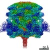
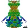
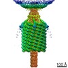
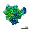
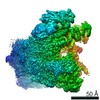
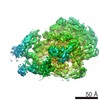
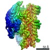
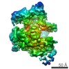
| Title | Myoviridae Phage XM1 Neck Region (12-fold) | |||||||||
 Map data Map data | 12 fold average for phage XM1 neck | |||||||||
 Sample Sample |
| |||||||||
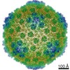
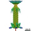
| Biological species |  Vibrio phage XM1 (virus) Vibrio phage XM1 (virus) | |||||||||
| Method |  single particle reconstruction / cryo EM / Resolution: 3.6 Å single particle reconstruction / cryo EM / Resolution: 3.6 Å | |||||||||
 Authors Authors | Wang Z / Klose T / Jiang W / Kuhn RJ | |||||||||
| Funding support |  United States, 1 items United States, 1 items
| |||||||||
 Citation Citation | Journal: Biorxiv / Year: 2021 Title: Structure of Vibrio phage XM1, a simple contractile DNA injection machine Authors: Wang Z / Fokine A / Guo X / Jiang W / Rossmann MG / Kuhn RJ / Luo ZH / Klose T | |||||||||
| History |
|
- Structure visualization
| Movie |
 Movie viewer Movie viewer |
|---|---|
| Structure viewer | EM map: SurfViewMolmilJmol/JSmol |
| Supplemental images |

- Downloads & links
-EMDB archive
| Map data | emd_22917.map.gz | 38.8 MB | EMDB map data format | |
|---|---|---|---|---|
| Header (meta data) | emd-22917-v30.xmlemd-22917.xml | 12.3 KB 12.3 KB | Display Display | EMDB header |
| FSC (resolution estimation) | emd_22917_fsc.xml | 18.1 KB | Display | FSC data file |
| Images | 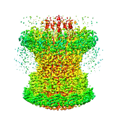 emd_22917.png emd_22917.png | 203.5 KB | ||
| Archive directory |  http://ftp.pdbj.org/pub/emdb/structures/EMD-22917ftp://ftp.pdbj.org/pub/emdb/structures/EMD-22917 http://ftp.pdbj.org/pub/emdb/structures/EMD-22917ftp://ftp.pdbj.org/pub/emdb/structures/EMD-22917 | HTTPS FTP |
-Related structure data
| Related structure data |  7klnMC  7kh1C  7kjkC  7kmxC M: atomic model generated by this map C: citing same article ( |
|---|---|
| Similar structure data |

-Links
| EMDB pages | EMDB (EBI/PDBe) / EMDataResource |
|---|
-Map
| File | Download / File: emd_22917.map.gz / Format: CCP4 / Size: 512 MB / Type: IMAGE STORED AS FLOATING POINT NUMBER (4 BYTES) | ||||||||||||||||||||||||||||||||||||||||||||||||||||||||||||||||||||
|---|---|---|---|---|---|---|---|---|---|---|---|---|---|---|---|---|---|---|---|---|---|---|---|---|---|---|---|---|---|---|---|---|---|---|---|---|---|---|---|---|---|---|---|---|---|---|---|---|---|---|---|---|---|---|---|---|---|---|---|---|---|---|---|---|---|---|---|---|---|
| Annotation | 12 fold average for phage XM1 neck | ||||||||||||||||||||||||||||||||||||||||||||||||||||||||||||||||||||
| Voxel size | X=Y=Z: 0.81 Å | ||||||||||||||||||||||||||||||||||||||||||||||||||||||||||||||||||||
| Density |
| ||||||||||||||||||||||||||||||||||||||||||||||||||||||||||||||||||||
| Symmetry | Space group: 1 | ||||||||||||||||||||||||||||||||||||||||||||||||||||||||||||||||||||
| Details | EMDB XML:
CCP4 map header:
| ||||||||||||||||||||||||||||||||||||||||||||||||||||||||||||||||||||
-Supplemental data
- Sample components
-Entire : Vibrio phage XM1
| Entire | Name: Vibrio phage XM1 (virus) |
|---|---|
| Components |
|
-Supramolecule #1: Vibrio phage XM1
| Supramolecule | Name: Vibrio phage XM1 / type: virus / ID: 1 / Parent: 0 / Macromolecule list: all / NCBI-ID: 2748688 / Sci species name: Vibrio phage XM1 / Virus type: VIRION / Virus isolate: OTHER / Virus enveloped: No / Virus empty: No |
|---|---|
| Host (natural) | Organism:  Vibrio rotiferianus (bacteria) Vibrio rotiferianus (bacteria) |
| Virus shell | Shell ID: 1 / Name: capsid / Diameter: 640.0 Å / T number (triangulation number): 7 |
-Macromolecule #1: Portal protein
| Macromolecule | Name: Portal protein / type: protein_or_peptide / ID: 1 / Number of copies: 12 / Enantiomer: LEVO |
|---|---|
| Source (natural) | Organism: Vibrio phage XM1 (virus) |
| Molecular weight | Theoretical: 47.018758 KDa |
| Sequence | String: MKFFDGVKDV LSGLINRRNS MARNRVSHRY LSDEEMRVMY KAGLMSKIIR LKAGYALNDT LKFESTQDQE IYKKRLSKHV KNATKFMLG FGRGVIVVFK NGDDLSKPLE RGVDPKLLKI RVFSGDIAKG NNPDNDLRSE RYYKPKNYTI KGHTIHWTRV V DFTYYMPS ...String: MKFFDGVKDV LSGLINRRNS MARNRVSHRY LSDEEMRVMY KAGLMSKIIR LKAGYALNDT LKFESTQDQE IYKKRLSKHV KNATKFMLG FGRGVIVVFK NGDDLSKPLE RGVDPKLLKI RVFSGDIAKG NNPDNDLRSE RYYKPKNYTI KGHTIHWTRV V DFTYYMPS ENELPDYYYG GMSESELIYE QFINDSVVQR ASGSIIEKAS TFVYKIKGYK QLIQAKKEED IIKYVSTCED GR SIYGGLI TDADDEVSTL TQSLTDLDKV DNVTLRRIAM VTGLGMTVLI GEQASGLNAS GEKERQGFQD TIENLQSDYL EDP LNRLAE IFQLGFIEFK DNQGQSANER VEYDKKAVDV AKVLWELGED YGAYLKDKDV VQADDWDNFW KEKDENSEVD ESLP LGDLF SSGDVNG |
-Macromolecule #2: Head completion protein, gp1
| Macromolecule | Name: Head completion protein, gp1 / type: protein_or_peptide / ID: 2 / Number of copies: 12 / Enantiomer: LEVO |
|---|---|
| Source (natural) | Organism: Vibrio phage XM1 (virus) |
| Molecular weight | Theoretical: 12.746998 KDa |
| Sequence | String: MALIDDFKAR FPNLDGSLVD ALVPVYENNY SCYYGGSYEN DCDKEAILLL IAHLVVTDPS YSGDESSSRA VASQSVGSVS VSFVAGSTG SDWTNWLNST RYGQLFLMVT SNNMGPSFA |
-Experimental details
-Structure determination
| Method | cryo EM |
|---|---|
 Processing Processing | single particle reconstruction |
| Aggregation state | particle |
-Sample preparation
| Buffer | pH: 7.5 / Details: 50 mM Tris, pH 7.5, 100 mM NaCl, 8 mM MgSO4 |
|---|---|
| Grid | Model: PELCO Ultrathin Carbon with Lacey Carbon / Material: COPPER / Mesh: 400 / Support film - Material: CARBON / Support film - topology: LACEY / Pretreatment - Type: GLOW DISCHARGE |
| Vitrification | Cryogen name: ETHANE / Chamber humidity: 80 % / Chamber temperature: 298 K / Instrument: GATAN CRYOPLUNGE 3 |
- Electron microscopy
| Microscope | FEI TITAN KRIOS |
|---|---|
| Electron beam | Acceleration voltage: 300 kV / Electron source: FIELD EMISSION GUN |
| Electron optics | Illumination mode: FLOOD BEAM / Imaging mode: BRIGHT FIELDBright-field microscopy / Cs: 2.7 mm / Nominal defocus max: 2.0 µm / Nominal defocus min: 0.5 µm |
| Sample stage | Specimen holder model: FEI TITAN KRIOS AUTOGRID HOLDER / Cooling holder cryogen: NITROGEN |
| Image recording | Film or detector model: DIRECT ELECTRON DE-16 (4k x 4k) / Detector mode: SUPER-RESOLUTION / Average electron dose: 25.0 e/Å2 |
| Experimental equipment |  Model: Titan Krios / Image courtesy: FEI Company |
-Image processing
| CTF correction | Software - Name: CTFFIND |
|---|---|
| Initial angle assignment | Type: RANDOM ASSIGNMENT |
| Final angle assignment | Type: PROJECTION MATCHING / Software - Name: jspr |
| Final reconstruction | Resolution.type: BY AUTHOR / Resolution: 3.6 Å / Resolution method: FSC 0.143 CUT-OFF / Software - Name: jspr / Number images used: 16015 |
| FSC plot (resolution estimation) |  |
-Atomic model buiding 1
| Refinement | Protocol: AB INITIO MODEL |
|---|---|
| Output model | PDB-7kln: |
