 Movie
Movie Controller


+ Open data

- Basic information
| Entry | Database: PDB / ID: 7kln | ||||||
|---|---|---|---|---|---|---|---|
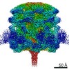
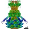
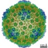
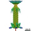
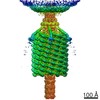
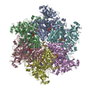
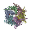
| Title | Myoviridae Phage XM1 Neck Region (12-fold) | ||||||
 Components Components |
| ||||||
 Keywords Keywords |  VIRAL PROTEIN / Myoviridae / Phage / Baseplate complex VIRAL PROTEIN / Myoviridae / Phage / Baseplate complex | ||||||
| Biological species |  Vibrio phage XM1 (virus) Vibrio phage XM1 (virus) | ||||||
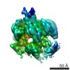
| Method | ELECTRON MICROSCOPY / single particle reconstruction / cryo EM / Resolution: 3.6 Å | ||||||
 Authors Authors | Wang, Z. / Klose, T. / Jiang, W. / Kuhn, R.J. | ||||||
| Funding support |  United States, 1items United States, 1items
| ||||||
 Citation Citation | Journal: Biorxiv / Year: 2021 Title: Structure of Vibrio phage XM1, a simple contractile DNA injection machine Authors: Wang, Z. / Fokine, A. / Guo, X. / Jiang, W. / Rossmann, M.G. / Kuhn, R.J. / Luo, Z.H. / Klose, T. | ||||||
| History |
|
- Structure visualization
| Movie |
 Movie viewer Movie viewer |
|---|---|
| Structure viewer | Molecule: MolmilJmol/JSmol |
- Downloads & links
-Download
| PDBx/mmCIF format | 7kln.cif.gz | 959.3 KB | Display | PDBx/mmCIF format |
|---|---|---|---|---|
| PDB format | pdb7kln.ent.gz | Display | PDB format | |
| PDBx/mmJSON format | 7kln.json.gz | Tree view | PDBx/mmJSON format | |
| Others |  Other downloads Other downloads |
-Validation report
| Arichive directory | https://data.pdbj.org/pub/pdb/validation_reports/kl/7klnftp://data.pdbj.org/pub/pdb/validation_reports/kl/7kln | HTTPS FTP |
|---|
-Related structure data
| Related structure data | 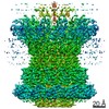 22917MC 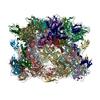 7kh1C 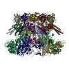 7kjkC  7kmxC M: map data used to model this data C: citing same article ( |
|---|---|
| Similar structure data |


-Links
 PDBj
PDBj- Assembly
| Deposited unit | 
|
|---|---|
| 1 |
|
-Components
| #1: Protein | Mass: 47018.758 Da / Num. of mol.: 12 / Source method: isolated from a natural source / Source: (natural) Vibrio phage XM1 (virus)#2: Protein | Mass: 12746.998 Da / Num. of mol.: 12 / Source method: isolated from a natural source / Source: (natural) Vibrio phage XM1 (virus) |
|---|
-Experimental details
-Experiment
| Experiment | Method: ELECTRON MICROSCOPY |
|---|---|
| EM experiment | Aggregation state: PARTICLE / 3D reconstruction method: single particle reconstruction |
- Sample preparation
| Component | Name: Vibrio phage XM1 / Type: VIRUS / Entity ID: all / Source: NATURAL |
|---|---|
| Molecular weight | Experimental value: NO |
| Source (natural) | Organism: Vibrio phage XM1 (virus) |
| Details of virus | Empty: NO / Enveloped: NO / Isolate: OTHER / Type: VIRION |
| Natural host | Organism: Vibrio rotiferianus |
| Virus shell | Name: capsid / Diameter: 640 nm / Triangulation number (T number): 7 |
| Buffer solution | pH: 7.5 / Details: 50 mM Tris, pH 7.5, 100 mM NaCl, 8 mM MgSO4 |
| Specimen | Embedding applied: NO / Shadowing applied: NO / Staining applied: NO / Vitrification applied: YES |
| Specimen support | Grid material: COPPER / Grid mesh size: 400 divisions/in. / Grid type: PELCO Ultrathin Carbon with Lacey Carbon |
| Vitrification | Instrument: GATAN CRYOPLUNGE 3 / Cryogen name: ETHANE / Humidity: 80 % / Chamber temperature: 298 K |
- Electron microscopy imaging
| Experimental equipment |  Model: Titan Krios / Image courtesy: FEI Company |
|---|---|
| Microscopy | Model: FEI TITAN KRIOS |
| Electron gun | Electron source: FIELD EMISSION GUN / Accelerating voltage: 300 kV / Illumination mode: FLOOD BEAM |
| Electron lens | Mode: BRIGHT FIELDBright-field microscopy / Nominal defocus max: 2000 nm / Nominal defocus min: 500 nm / Cs: 2.7 mm / Alignment procedure: COMA FREE |
| Specimen holder | Cryogen: NITROGEN / Specimen holder model: FEI TITAN KRIOS AUTOGRID HOLDER |
| Image recording | Electron dose: 25 e/Å2 / Detector mode: SUPER-RESOLUTION / Film or detector model: DIRECT ELECTRON DE-16 (4k x 4k) |
- Processing
| EM software |
| ||||||||||||||||||
|---|---|---|---|---|---|---|---|---|---|---|---|---|---|---|---|---|---|---|---|
| CTF correction | Type: NONE | ||||||||||||||||||
| 3D reconstruction | Resolution: 3.6 Å / Resolution method: FSC 0.143 CUT-OFF / Num. of particles: 16015 / Symmetry type: POINT | ||||||||||||||||||
| Atomic model building | Protocol: AB INITIO MODEL |
