 Movie
Movie Controller


+ Open data

- Basic information
| Entry | Database: EMDB / ID: EMD-20673 | |||||||||
|---|---|---|---|---|---|---|---|---|---|---|
| Title | Cryo-EM Structure of Helical Lipoprotein Lipase | |||||||||
 Map data Map data | Helical Lipoprotein Lipase | |||||||||
 Sample Sample |
| |||||||||
| Function / homology |  Function and homology information Function and homology informationAssembly of active LPL and LIPC lipase complexes / positive regulation of chemokine production => GO:0032722 /  lipoprotein lipase / lipoprotein lipase activity / positive regulation of tumor necrosis factor production => GO:0032760 / positive regulation of sequestering of triglyceride / low-density lipoprotein particle mediated signaling / chylomicron remodeling / positive regulation of cholesterol storage / phospholipase A1 ...Assembly of active LPL and LIPC lipase complexes / positive regulation of chemokine production => GO:0032722 / lipoprotein lipase / lipoprotein lipase activity / positive regulation of tumor necrosis factor production => GO:0032760 / positive regulation of sequestering of triglyceride / low-density lipoprotein particle mediated signaling / chylomicron remodeling / positive regulation of cholesterol storage / phospholipase A1 / phosphatidylserine 1-acylhydrolase activity / 1-acyl-2-lysophosphatidylserine acylhydrolase activity / phospholipase A1 activity / phospholipase activity / triglyceride catabolic process / lipase activity / very-low-density lipoprotein particle remodeling / positive regulation of macrophage derived foam cell differentiation / chylomicron / cellular response to nutrient / very-low-density lipoprotein particle / triglyceride lipase activity / positive regulation of chemokine (C-X-C motif) ligand 2 production / heparan sulfate proteoglycan binding / triglyceride homeostasis / cellular response to fatty acid / triglyceride metabolic process / lipoprotein particle binding / apolipoprotein binding / positive regulation of fat cell differentiation / phospholipid metabolic process / response to glucose / lipid catabolic process / positive regulation of interleukin-1 beta production / cholesterol homeostasis / response to bacterium / fatty acid biosynthetic process / positive regulation of inflammatory response / positive regulation of interleukin-6 production / heparin binding / signaling receptor binding / calcium ion binding / cell surface / protein homodimerization activity / extracellular space / plasma membrane lipoprotein lipase / lipoprotein lipase activity / positive regulation of tumor necrosis factor production => GO:0032760 / positive regulation of sequestering of triglyceride / low-density lipoprotein particle mediated signaling / chylomicron remodeling / positive regulation of cholesterol storage / phospholipase A1 ...Assembly of active LPL and LIPC lipase complexes / positive regulation of chemokine production => GO:0032722 / lipoprotein lipase / lipoprotein lipase activity / positive regulation of tumor necrosis factor production => GO:0032760 / positive regulation of sequestering of triglyceride / low-density lipoprotein particle mediated signaling / chylomicron remodeling / positive regulation of cholesterol storage / phospholipase A1 / phosphatidylserine 1-acylhydrolase activity / 1-acyl-2-lysophosphatidylserine acylhydrolase activity / phospholipase A1 activity / phospholipase activity / triglyceride catabolic process / lipase activity / very-low-density lipoprotein particle remodeling / positive regulation of macrophage derived foam cell differentiation / chylomicron / cellular response to nutrient / very-low-density lipoprotein particle / triglyceride lipase activity / positive regulation of chemokine (C-X-C motif) ligand 2 production / heparan sulfate proteoglycan binding / triglyceride homeostasis / cellular response to fatty acid / triglyceride metabolic process / lipoprotein particle binding / apolipoprotein binding / positive regulation of fat cell differentiation / phospholipid metabolic process / response to glucose / lipid catabolic process / positive regulation of interleukin-1 beta production / cholesterol homeostasis / response to bacterium / fatty acid biosynthetic process / positive regulation of inflammatory response / positive regulation of interleukin-6 production / heparin binding / signaling receptor binding / calcium ion binding / cell surface / protein homodimerization activity / extracellular space / plasma membraneSimilarity search - Function | |||||||||
| Biological species |  Bos taurus (cattle) / Bovine (cattle) Bos taurus (cattle) / Bovine (cattle) | |||||||||
| Method | helical reconstruction / cryo EM / Resolution: 3.8 Å | |||||||||
 Authors Authors | Gunn KH / Wang F / Egelman EH / Neher SB | |||||||||
| Funding support |  United States, 2 items United States, 2 items
| |||||||||
 Citation Citation | Journal: Proc Natl Acad Sci U S A / Year: 2020 Title: The structure of helical lipoprotein lipase reveals an unexpected twist in lipase storage. Authors: Kathryn H Gunn / Benjamin S Roberts / Fengbin Wang / Joshua D Strauss / Mario J Borgnia / Edward H Egelman / Saskia B Neher / Abstract: Lipases are enzymes necessary for the proper distribution and utilization of lipids in the human body. Lipoprotein lipase (LPL) is active in capillaries, where it plays a crucial role in preventing ...Lipases are enzymes necessary for the proper distribution and utilization of lipids in the human body. Lipoprotein lipase (LPL) is active in capillaries, where it plays a crucial role in preventing dyslipidemia by hydrolyzing triglycerides from packaged lipoproteins. Thirty years ago, the existence of a condensed and inactive LPL oligomer was proposed. Although recent work has shed light on the structure of the LPL monomer, the inactive oligomer remained opaque. Here we present a cryo-EM reconstruction of a helical LPL oligomer at 3.8-Å resolution. Helix formation is concentration-dependent, and helices are composed of inactive dihedral LPL dimers. Heparin binding stabilizes LPL helices, and the presence of substrate triggers helix disassembly. Superresolution fluorescent microscopy of endogenous LPL revealed that LPL adopts a filament-like distribution in vesicles. Mutation of one of the helical LPL interaction interfaces causes loss of the filament-like distribution. Taken together, this suggests that LPL is condensed into its inactive helical form for storage in intracellular vesicles. | |||||||||
| History |
|
- Structure visualization
| Movie |
Movie viewer |
|---|---|
| Structure viewer | EM map: SurfViewMolmilJmol/JSmol |
| Supplemental images |

- Downloads & links
-EMDB archive
| Map data | emd_20673.map.gz | 97.1 MB | EMDB map data format | |
|---|---|---|---|---|
| Header (meta data) | emd-20673-v30.xmlemd-20673.xml | 14.2 KB 14.2 KB | Display Display | EMDB header |
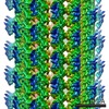
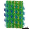
| Images | 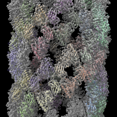 emd_20673.png emd_20673.png | 323.6 KB | ||
| Archive directory |  http://ftp.pdbj.org/pub/emdb/structures/EMD-20673ftp://ftp.pdbj.org/pub/emdb/structures/EMD-20673 http://ftp.pdbj.org/pub/emdb/structures/EMD-20673ftp://ftp.pdbj.org/pub/emdb/structures/EMD-20673 | HTTPS FTP |
-Related structure data
| Related structure data |  6u7mMC M: atomic model generated by this map C: citing same article ( |
|---|---|
| Similar structure data |


-Links
| EMDB pages | EMDB (EBI/PDBe) / EMDataResource |
|---|
-Map
| File | Download / File: emd_20673.map.gz / Format: CCP4 / Size: 216 MB / Type: IMAGE STORED AS FLOATING POINT NUMBER (4 BYTES) | ||||||||||||||||||||||||||||||||||||||||||||||||||||||||||||
|---|---|---|---|---|---|---|---|---|---|---|---|---|---|---|---|---|---|---|---|---|---|---|---|---|---|---|---|---|---|---|---|---|---|---|---|---|---|---|---|---|---|---|---|---|---|---|---|---|---|---|---|---|---|---|---|---|---|---|---|---|---|
| Annotation | Helical Lipoprotein Lipase | ||||||||||||||||||||||||||||||||||||||||||||||||||||||||||||
| Voxel size | X=Y=Z: 0.9317 Å | ||||||||||||||||||||||||||||||||||||||||||||||||||||||||||||
| Density |
| ||||||||||||||||||||||||||||||||||||||||||||||||||||||||||||
| Symmetry | Space group: 1 | ||||||||||||||||||||||||||||||||||||||||||||||||||||||||||||
| Details | EMDB XML:
CCP4 map header:
| ||||||||||||||||||||||||||||||||||||||||||||||||||||||||||||
-Supplemental data
- Sample components
-Entire : filament of lipoprotein lipase
| Entire | Name: filament of lipoprotein lipase |
|---|---|
| Components |
|
-Supramolecule #1: filament of lipoprotein lipase
| Supramolecule | Name: filament of lipoprotein lipase / type: complex / ID: 1 / Parent: 0 / Macromolecule list: all |
|---|---|
| Source (natural) | Organism: Bos taurus (cattle) |
-Macromolecule #1: Lipoprotein lipase
| Macromolecule | Name: Lipoprotein lipase / type: protein_or_peptide / ID: 1 / Number of copies: 30 / Enantiomer: LEVO / EC number: lipoprotein lipase |
|---|---|
| Source (natural) | Organism: Bovine (cattle) |
| Molecular weight | Theoretical: 53.448789 KDa |
| Sequence | String: MESKALLLLA LSVCLQSLTV SRGGLVAADR ITGGKDFRDI ESKFALRTPE DTAEDTCHLI PGVTESVANC HFNHSSKTFV VIHGWTVTG MYESWVPKLV AALYKREPDS NVIVVDWLSR AQQHYPVSAG YTKLVGQDVA KFMNWMADEF NYPLGNVHLL G YSLGAHAA ...String: MESKALLLLA LSVCLQSLTV SRGGLVAADR ITGGKDFRDI ESKFALRTPE DTAEDTCHLI PGVTESVANC HFNHSSKTFV VIHGWTVTG MYESWVPKLV AALYKREPDS NVIVVDWLSR AQQHYPVSAG YTKLVGQDVA KFMNWMADEF NYPLGNVHLL G YSLGAHAA GIAGSLTNKK VNRITGLDPA GPNFEYAEAP SRLSPDDADF VDVLHTFTRG SPGRSIGIQK PVGHVDIYPN GG TFQPGCN IGEALRVIAE RGLGDVDQLV KCSHERSVHL FIDSLLNEEN PSKAYRCNSK EAFEKGLCLS CRKNRCNNMG YEI NKVRAK RSSKMYLKTR SQMPYKVFHY QVKIHFSGTE SNTYTNQAFE ISLYGTVAES ENIPFTLPEV STNKTYSFLL YTEV DIGEL LMLKLKWISD SYFSWSNWWS SPGFDIGKIR VKAGETQKKV IFCSREKMSY LQKGKSPVIF VKCHDKSLNR KSG |
-Experimental details
-Structure determination
| Method | cryo EM |
|---|---|
 Processing Processing | helical reconstruction |
| Aggregation state | filament |
-Sample preparation
| Concentration | 0.35 mg/mL | |||||||||||||||
|---|---|---|---|---|---|---|---|---|---|---|---|---|---|---|---|---|
| Buffer | pH: 8 Component:
| |||||||||||||||
| Grid | Pretreatment - Type: PLASMA CLEANING / Pretreatment - Atmosphere: AIR / Pretreatment - Pressure: 0.038 kPa / Details: 15 mA current | |||||||||||||||
| Vitrification | Cryogen name: ETHANE / Chamber humidity: 98 % / Chamber temperature: 295 K / Instrument: LEICA EM GP / Details: Blot 3 seconds before plunging. |
- Electron microscopy
| Microscope | FEI TALOS ARCTICA |
|---|---|
| Electron beam | Acceleration voltage: 200 kV / Electron source: FIELD EMISSION GUN |
| Electron optics | C2 aperture diameter: 70.0 µm / Illumination mode: FLOOD BEAM / Imaging mode: BRIGHT FIELDBright-field microscopy / Cs: 2.7 mm / Nominal magnification: 45000 |
| Sample stage | Cooling holder cryogen: NITROGEN |
| Image recording | Film or detector model: GATAN K2 SUMMIT (4k x 4k) / Detector mode: COUNTING / Number real images: 1764 / Average exposure time: 12.8 sec. / Average electron dose: 46.6 e/Å2 |
| Experimental equipment |  Model: Talos Arctica / Image courtesy: FEI Company |
-Image processing
| Segment selection | Number selected: 157128 Software:
| ||||||
|---|---|---|---|---|---|---|---|
| CTF correction | Software - Name: CTFFIND (ver. 4) | ||||||
| Startup model | Type of model: OTHER Details: 127 A radius featureless cylinder generated in RELION | ||||||
| Final angle assignment | Type: NOT APPLICABLE / Software - Name: RELION (ver. 3.0) | ||||||
| Final reconstruction | Number classes used: 1 Applied symmetry - Helical parameters - Δz: 10.88 Å Applied symmetry - Helical parameters - Δ&Phi: 130.05 ° Applied symmetry - Helical parameters - Axial symmetry: C1 (asymmetric) Algorithm: BACK PROJECTION / Resolution.type: BY AUTHOR / Resolution: 3.8 Å / Resolution method: OTHER / Software - Name: RELION (ver. 3.0) Details: model: map FSC 0.38 cutoff; map: map FSC 0.143 cutoff Number images used: 108911 |

