 Movie
Movie Controller

[English] 日本語
 Yorodumi
- EMDB-18918: Subtomogram average of the Vaccinia virus (WR) A4/A10 palisade tr... -
+ Open data

- Basic information
| Entry |  | |||||||||
|---|---|---|---|---|---|---|---|---|---|---|
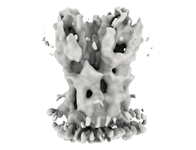
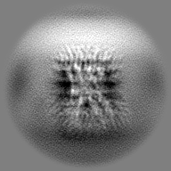
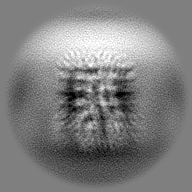
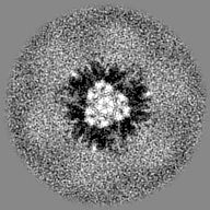
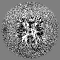
| Title | Subtomogram average of the Vaccinia virus (WR) A4/A10 palisade trimer in mature virions | |||||||||
 Map data Map data | ||||||||||
 Sample Sample |
| |||||||||
 Keywords Keywords | Palisade / A10 / A4 / p4a /  Vaccinia / VIRAL PROTEIN Vaccinia / VIRAL PROTEIN | |||||||||
| Function / homology |  Function and homology informationvirion component / host cell endoplasmic reticulum-Golgi intermediate compartment membrane / structural molecule activity / membrane Function and homology informationvirion component / host cell endoplasmic reticulum-Golgi intermediate compartment membrane / structural molecule activity / membraneSimilarity search - Function | |||||||||
| Biological species |  Vaccinia virus WR Vaccinia virus WR | |||||||||
| Method | subtomogram averaging / cryo EM / Resolution: 9.7 Å | |||||||||
 Authors Authors | Calcraft T / Hernandez-Gonzalez M / Nans A / Rosenthal PB / Way M | |||||||||
| Funding support |  United Kingdom, 1 items United Kingdom, 1 items
| |||||||||
 Citation Citation | Journal: mBio / Year: 2024 Title: Palisade structure in intact vaccinia virions. Authors: Miguel Hernandez-Gonzalez / Thomas Calcraft / Andrea Nans / Peter B Rosenthal / Michael Way / Abstract: Vaccinia virus assembly in the cytoplasm of infected cells involves the formation of a biconcave viral core inside the maturing viral particle. The boundary of the core is defined by a ...Vaccinia virus assembly in the cytoplasm of infected cells involves the formation of a biconcave viral core inside the maturing viral particle. The boundary of the core is defined by a pseudohexagonal palisade layer, composed of trimers projecting from an inner wall. To understand the assembly of this complex core architecture, we obtained a subnanometer structure of the palisade trimer by cryo-electron tomography and subtomogram averaging of purified intact virions. Using AlphaFold2 structure predictions, we determined that the palisade is formed from trimers of the proteolytically processed form of the viral protein A10. In addition, we found that each A10 protomer associates with an α-helix (residues 24-66) of A4. Cellular localization assays outside the context of infection demonstrate that the A4 N-terminus is necessary and sufficient to interact with A10. The interaction between A4 and A10 provides insights into how the palisade layer might become tightly associated with the viral membrane during virion maturation. Reconstruction of the palisade layer reveals that, despite local hexagonal ordering, the A10/A4 trimers are widely spaced, suggesting that additional components organize the lattice. This spacing would, however, allow the adoption of the characteristic biconcave shape of the viral core. Finally, we also found that the palisade incorporates multiple copies of a hexameric portal structure. We suggest that these portals are formed by E6, a viral protein that is essential for virion assembly and required to release viral mRNA from the core early in infection.IMPORTANCEPoxviruses such as variola virus (smallpox) and monkeypox cause diseases in humans. Other poxviruses, including vaccinia and modified vaccinia Ankara, are used as vaccine vectors. Given their importance, a greater structural understanding of poxvirus virions is needed. We now performed cryo-electron tomography of purified intact vaccinia virions to study the structure of the palisade, a protein lattice that defines the viral core boundary. We identified the main viral proteins that form the palisade and their interaction surfaces and provided new insights into the organization of the viral core. | |||||||||
| History |
|
- Structure visualization
| Supplemental images |
|---|
- Downloads & links
-EMDB archive
| Map data | emd_18918.map.gz | 2.3 MB | EMDB map data format | |
|---|---|---|---|---|
| Header (meta data) | emd-18918-v30.xmlemd-18918.xml | 17.7 KB 17.7 KB | Display Display | EMDB header |
| FSC (resolution estimation) | emd_18918_fsc.xml | 6.9 KB | Display | FSC data file |
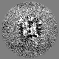
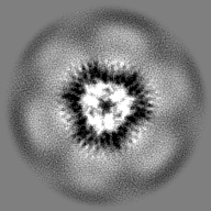
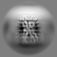
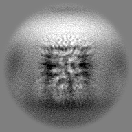
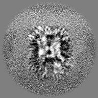
| Images |  emd_18918.png emd_18918.png | 71 KB | ||
| Masks | emd_18918_msk_1.map | 27 MB | Mask map | |
| Filedesc metadata | emd-18918.cif.gz | 6.5 KB | ||
| Others | emd_18918_half_map_1.map.gzemd_18918_half_map_2.map.gz | 13.3 MB 13.3 MB | ||
| Archive directory |  http://ftp.pdbj.org/pub/emdb/structures/EMD-18918ftp://ftp.pdbj.org/pub/emdb/structures/EMD-18918 http://ftp.pdbj.org/pub/emdb/structures/EMD-18918ftp://ftp.pdbj.org/pub/emdb/structures/EMD-18918 | HTTPS FTP |
-Related structure data
| Related structure data | 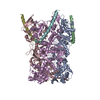 8r5iMC M: atomic model generated by this map C: citing same article ( |
|---|---|
| Similar structure data |


-Links
| EMDB pages | EMDB (EBI/PDBe) / EMDataResource |
|---|
-Map
| File | Download / File: emd_18918.map.gz / Format: CCP4 / Size: 27 MB / Type: IMAGE STORED AS FLOATING POINT NUMBER (4 BYTES) | ||||||||||||||||||||
|---|---|---|---|---|---|---|---|---|---|---|---|---|---|---|---|---|---|---|---|---|---|
| Voxel size | X=Y=Z: 1.56 Å | ||||||||||||||||||||
| Density |
| ||||||||||||||||||||
| Symmetry | Space group: 1 | ||||||||||||||||||||
| Details | EMDB XML:
|
-Supplemental data
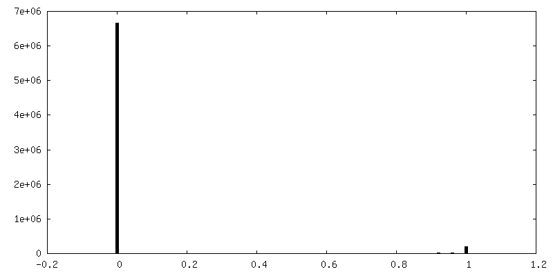
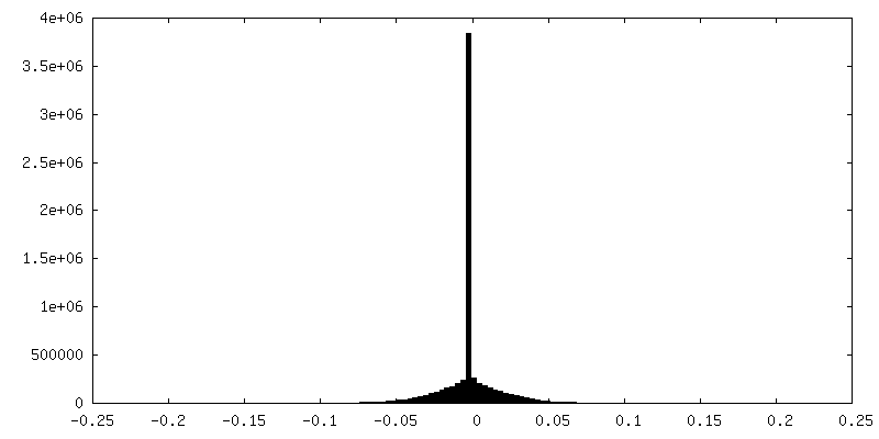
-Mask #1
| File | emd_18918_msk_1.map | ||||||||||||
|---|---|---|---|---|---|---|---|---|---|---|---|---|---|
| Projections & Slices |
| ||||||||||||
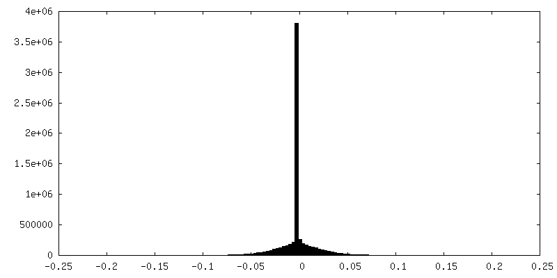
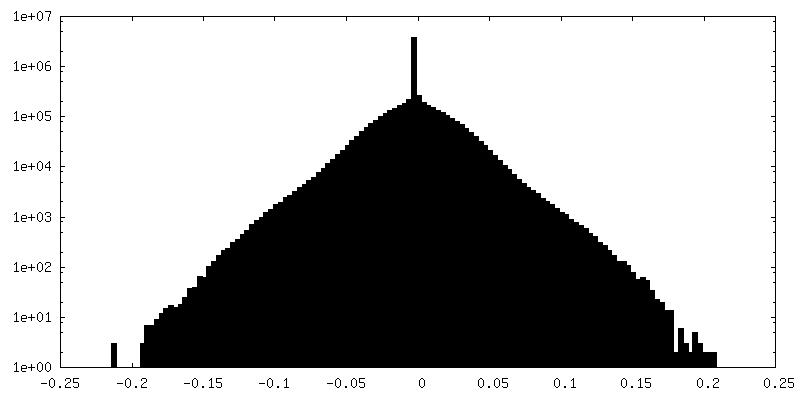
| Density Histograms |
 Z
Z Y
Y X
X


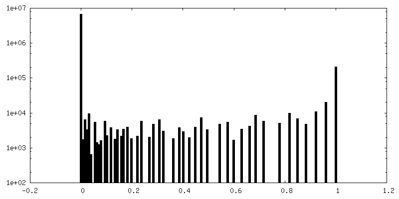
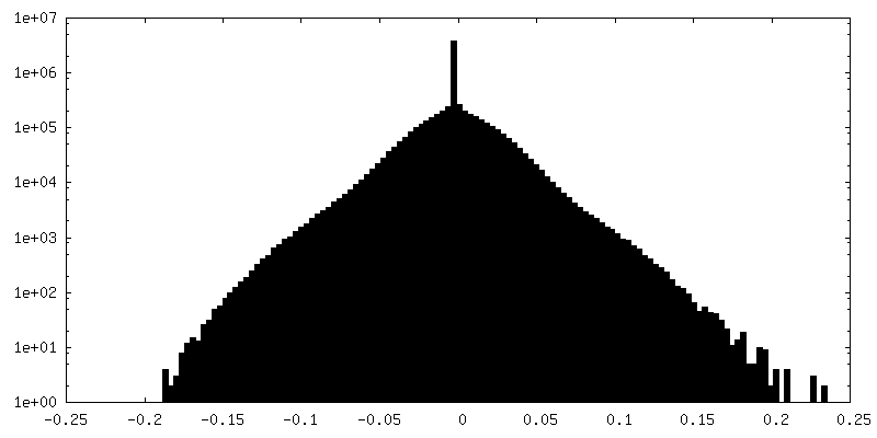
-Half map: #1
| File | emd_18918_half_map_1.map | ||||||||||||
|---|---|---|---|---|---|---|---|---|---|---|---|---|---|
| Projections & Slices |
| ||||||||||||
| Density Histograms |

-Half map: #2
| File | emd_18918_half_map_2.map | ||||||||||||
|---|---|---|---|---|---|---|---|---|---|---|---|---|---|
| Projections & Slices |
| ||||||||||||
| Density Histograms |

- Sample components
-Entire : Vaccinia virus WR
| Entire | Name: Vaccinia virus WR |
|---|---|
| Components |
|
-Supramolecule #1: Vaccinia virus WR
| Supramolecule | Name: Vaccinia virus WR / type: virus / ID: 1 / Parent: 0 / Macromolecule list: all / Details: A36-YdF / NCBI-ID: 10254 / Sci species name: Vaccinia virus WR / Virus type: VIRION / Virus isolate: STRAIN / Virus enveloped: Yes / Virus empty: No |
|---|---|
| Host (natural) | Organism:  Homo sapiens (human) Homo sapiens (human) |
-Macromolecule #1: Core protein A10
| Macromolecule | Name: Core protein A10 / type: protein_or_peptide / ID: 1 / Number of copies: 3 / Enantiomer: LEVO |
|---|---|
| Source (natural) | Organism: Vaccinia virus WR |
| Molecular weight | Theoretical: 71.073477 KDa |
| Recombinant expression | Organism: Homo sapiens (human) |
| Sequence | String: MMPIKSIVTL DQLEDSEYLF RIVSTVLPHL CLDYKVCDQL KTTFVHPFDI LLNNSLGSVT KQDELQAAIS KLGINYLIDT TSRELKLFN VTLNAGNIDI INTPINISSE TNPIINTHSF YDLPPFTQHL LNIRLTDTEY RARFIGGYIK PDGSDSMDVL A EKKYPDLN ...String: MMPIKSIVTL DQLEDSEYLF RIVSTVLPHL CLDYKVCDQL KTTFVHPFDI LLNNSLGSVT KQDELQAAIS KLGINYLIDT TSRELKLFN VTLNAGNIDI INTPINISSE TNPIINTHSF YDLPPFTQHL LNIRLTDTEY RARFIGGYIK PDGSDSMDVL A EKKYPDLN FDNTYLFNIL YKDVINAPIK EFKAKIVNGV LSRQDFDNLI GVRQYITIQD RPRFDDAYNI ADAARHYGVN LN TLPLPNV DLTTMPTYKH LIMFEQYFIY TYDRVDIYYN GNKMLFDDEI INFTISMRYQ SLIPRLVDFF PDIPVNNNIV LHT RDPQNA AVNVTVALPN VQFVDINRNN KFFINFFNLL AKEQRSTAIK VTKSMFWDGM DYEEYKSKNL QDMMFINSTC YVFG LYNHN NTTYCSILSD IISAEKTPIR VCLLPRVVGG KTVTNLISET LKSISSMTIR EFPRKDKSIM HIGLSETGFM RFFQL LRLM ADKPHETAIK EVVMAYVGIK LGDKGSPYYI RKESYQDFIY LLFASMGFKV TTRRSIMGSN NISIISIRPR VTKQYI VAT LMKTSCSKNE AEKLITSAFD LLNFMVSVSD FRDYQSYRQY RNYCPRYFYA G UniProtKB: Major core protein OPG136 precursor |
-Macromolecule #2: Core protein A4
| Macromolecule | Name: Core protein A4 / type: protein_or_peptide / ID: 2 / Number of copies: 3 / Enantiomer: LEVO |
|---|---|
| Source (natural) | Organism: Vaccinia virus WR |
| Molecular weight | Theoretical: 30.95235 KDa |
| Recombinant expression | Organism: Homo sapiens (human) |
| Sequence | String: MDFFNKFSQG LAESSTPKSS IYYSEEKDPD TKKDEAIEIG LKSQESYYQR QLREQLARDN MTVASRQPIQ PLQPTIHITP QPVPTATPA PILLPSSTVP TPKPRQQTNT SSDMSNLFDW LSEDTDAPAS SLLPALTPSN AVQDIISKFN KDQKTTTPPS T QPSQTLPT ...String: MDFFNKFSQG LAESSTPKSS IYYSEEKDPD TKKDEAIEIG LKSQESYYQR QLREQLARDN MTVASRQPIQ PLQPTIHITP QPVPTATPA PILLPSSTVP TPKPRQQTNT SSDMSNLFDW LSEDTDAPAS SLLPALTPSN AVQDIISKFN KDQKTTTPPS T QPSQTLPT TTCTQQSDGN ISCTTPTVTP PQPPIVATVC TPTPTGGTVC TTAQQNPNPG AASQQNLDDM ALKDLMSNVE RD MHQLQAE TNDLVTNVYD AREYTRRAID QILQLVKGFE RFQK UniProtKB: 39kDa core protein OPG130 |
-Experimental details
-Structure determination
| Method | cryo EM |
|---|---|
 Processing Processing | subtomogram averaging |
| Aggregation state | particle |
-Sample preparation
| Buffer | pH: 7.5 |
|---|---|
| Grid | Model: Quantifoil R2/2 / Material: COPPER / Mesh: 300 / Pretreatment - Type: GLOW DISCHARGE / Pretreatment - Time: 40 sec. / Details: 45mA |
| Vitrification | Cryogen name: ETHANE / Chamber humidity: 95 % / Chamber temperature: 295 K / Instrument: FEI VITROBOT MARK IV |
- Electron microscopy
| Microscope | FEI TITAN KRIOS |
|---|---|
| Electron beam | Acceleration voltage: 300 kV / Electron source: FIELD EMISSION GUN |
| Electron optics | Illumination mode: FLOOD BEAM / Imaging mode: BRIGHT FIELDBright-field microscopy / Cs: 2.7 mm / Nominal defocus max: 5.0 µm / Nominal defocus min: 2.0 µm |
| Specialist optics | Energy filter - Name: TFS Selectris / Energy filter - Slit width: 10 eV |
| Sample stage | Specimen holder model: FEI TITAN KRIOS AUTOGRID HOLDER / Cooling holder cryogen: NITROGEN |
| Image recording | Film or detector model: TFS FALCON 4i (4k x 4k) / Average electron dose: 2.4 e/Å2 |
| Experimental equipment |  Model: Titan Krios / Image courtesy: FEI Company |
-Image processing
| Extraction | Number tomograms: 98 / Number images used: 623174 / Software - Name: Dynamo |
|---|---|
| Final angle assignment | Type: MAXIMUM LIKELIHOOD / Software - Name: RELION (ver. 4.0) |
| Final reconstruction | Applied symmetry - Point group: C3 (3 fold cyclic) / Algorithm: FOURIER SPACE / Resolution.type: BY AUTHOR / Resolution: 9.7 Å / Resolution method: FSC 0.143 CUT-OFF / Software - Name: RELION (ver. 4.0) / Number subtomograms used: 123492 |
| FSC plot (resolution estimation) | 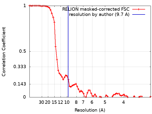 |
-Atomic model buiding 1
| Initial model | Chain - Source name: AlphaFold / Chain - Initial model type: in silico model |
|---|---|
| Refinement | Space: REAL / Protocol: FLEXIBLE FIT |
| Output model | PDB-8r5i: |
