- EMDB-14872: Plastocyanin bound to PSI of Chlamydomonas reinhardtii -
+
Open data
ID or keywords:
Loading...
-
Basic information
Entry
Database: EMDB / ID: EMD-14872
Title
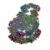
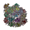
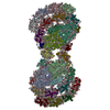
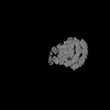
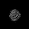
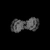
Plastocyanin bound to PSI of Chlamydomonas reinhardtii
Map data
Sample
Organelle or cellular component: Photosystem I dimer of Chlamydomonas reinhardtii
Protein or peptide: Plastocyanin, chloroplastic
Ligand: COPPER (II) ION
Function / homology
Function and homology information
electron transporter, transferring electrons from cytochrome b6/f complex of photosystem II activity / chloroplast thylakoid lumen / chloroplast thylakoid membrane / copper ion binding Similarity search - Function
Plastocyanin / Blue (type 1) copper protein, plastocyanin-type / Blue (type 1) copper domain / Copper binding proteins, plastocyanin/azurin family / Blue (type 1) copper protein, binding site / Type-1 copper (blue) proteins signature. / Cupredoxin Similarity search - Domain/homology
German Federal Ministry for Education and Research
NRW 28 313-WO44A
Germany
German-Israeli Foundation for Research and Development
G-1483- 207/2018
Germany
Citation
Journal: Nat Plants / Year: 2022 Title: Algal photosystem I dimer and high-resolution model of PSI-plastocyanin complex. Authors: Andreas Naschberger / Laura Mosebach / Victor Tobiasson / Sebastian Kuhlgert / Martin Scholz / Annemarie Perez-Boerema / Thi Thu Hoai Ho / André Vidal-Meireles / Yuichiro Takahashi / ...Authors: Andreas Naschberger / Laura Mosebach / Victor Tobiasson / Sebastian Kuhlgert / Martin Scholz / Annemarie Perez-Boerema / Thi Thu Hoai Ho / André Vidal-Meireles / Yuichiro Takahashi / Michael Hippler / Alexey Amunts / Abstract: Photosystem I (PSI) enables photo-electron transfer and regulates photosynthesis in the bioenergetic membranes of cyanobacteria and chloroplasts. Being a multi-subunit complex, its macromolecular ...Photosystem I (PSI) enables photo-electron transfer and regulates photosynthesis in the bioenergetic membranes of cyanobacteria and chloroplasts. Being a multi-subunit complex, its macromolecular organization affects the dynamics of photosynthetic membranes. Here we reveal a chloroplast PSI from the green alga Chlamydomonas reinhardtii that is organized as a homodimer, comprising 40 protein subunits with 118 transmembrane helices that provide scaffold for 568 pigments. Cryogenic electron microscopy identified that the absence of PsaH and Lhca2 gives rise to a head-to-head relative orientation of the PSI-light-harvesting complex I monomers in a way that is essentially different from the oligomer formation in cyanobacteria. The light-harvesting protein Lhca9 is the key element for mediating this dimerization. The interface between the monomers is lacking PsaH and thus partially overlaps with the surface area that would bind one of the light-harvesting complex II complexes in state transitions. We also define the most accurate available PSI-light-harvesting complex I model at 2.3 Å resolution, including a flexibly bound electron donor plastocyanin, and assign correct identities and orientations to all the pigments, as well as 621 water molecules that affect energy transfer pathways.
In the structure databanks used in Yorodumi, some data are registered as the other names, "COVID-19 virus" and "2019-nCoV". Here are the details of the virus and the list of structure data.
Jan 31, 2019. EMDB accession codes are about to change! (news from PDBe EMDB page)
EMDB accession codes are about to change! (news from PDBe EMDB page)
The allocation of 4 digits for EMDB accession codes will soon come to an end. Whilst these codes will remain in use, new EMDB accession codes will include an additional digit and will expand incrementally as the available range of codes is exhausted. The current 4-digit format prefixed with “EMD-” (i.e. EMD-XXXX) will advance to a 5-digit format (i.e. EMD-XXXXX), and so on. It is currently estimated that the 4-digit codes will be depleted around Spring 2019, at which point the 5-digit format will come into force.
The EM Navigator/Yorodumi systems omit the EMD- prefix.
Related info.:Q: What is EMD? / ID/Accession-code notation in Yorodumi/EM Navigator
Yorodumi is a browser for structure data from EMDB, PDB, SASBDB, etc.
This page is also the successor to EM Navigator detail page, and also detail information page/front-end page for Omokage search.
The word "yorodu" (or yorozu) is an old Japanese word meaning "ten thousand". "mi" (miru) is to see.
Related info.:EMDB / PDB / SASBDB / Comparison of 3 databanks / Yorodumi Search / Aug 31, 2016. New EM Navigator & Yorodumi / Yorodumi Papers / Jmol/JSmol / Function and homology information / Changes in new EM Navigator and Yorodumi
 Movie
Movie Controller


 Open data
 Basic information
 Map data
 Sample
 Function and homology information

 Chlamydomonas reinhardtii (plant)
 Authors
 Sweden,
  Germany, 4 items
  Citation

 Structure visualization
 Downloads & links
 emd_14872.png
 http://ftp.pdbj.org/pub/emdb/structures/EMD-14872
 Z (Sec.)
 Y (Row.)
 X (Col.)


 Sample components
 Processing
 Electron microscopy

