ムービー
ムービー コントローラー


+ データを開く

- 基本情報
基本情報
| 登録情報 | データベース: EMDB / ID: EMD-11079 | ||||||||||||
|---|---|---|---|---|---|---|---|---|---|---|---|---|---|
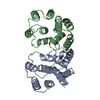
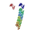
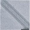
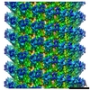
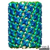
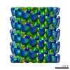
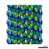
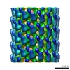
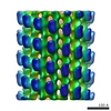
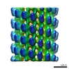
| タイトル | Helical reconstruction of influenza A virus M1 in complex with nucleic acid | ||||||||||||
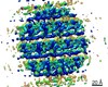
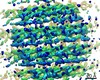
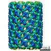
 マップデータ マップデータ | symmetrised helical reconstruction map | ||||||||||||
 試料 試料 |
| ||||||||||||
 キーワード キーワード | M1 /  Matrix protein (基質タンパク質) / Influenza virus (オルトミクソウイルス科) / Assembly / ribonucleoprotein complex (核タンパク質) / VIRAL PROTEIN (ウイルスタンパク質) Matrix protein (基質タンパク質) / Influenza virus (オルトミクソウイルス科) / Assembly / ribonucleoprotein complex (核タンパク質) / VIRAL PROTEIN (ウイルスタンパク質) | ||||||||||||
| 機能・相同性 |  機能・相同性情報 機能・相同性情報Assembly of Viral Components at the Budding Site / Influenza Infection / Fusion of the Influenza Virion to the Host Cell Endosome / Release / 出芽 / Packaging of Eight RNA Segments / Uncoating of the Influenza Virion / Entry of Influenza Virion into Host Cell via Endocytosis / Viral RNP Complexes in the Host Cell Nucleus / NEP/NS2 Interacts with the Cellular Export Machinery ...Assembly of Viral Components at the Budding Site / Influenza Infection / Fusion of the Influenza Virion to the Host Cell Endosome / Release / 出芽 / Packaging of Eight RNA Segments / Uncoating of the Influenza Virion / Entry of Influenza Virion into Host Cell via Endocytosis / Viral RNP Complexes in the Host Cell Nucleus / NEP/NS2 Interacts with the Cellular Export Machinery / Viral mRNA Translation / viral budding from plasma membrane / structural constituent of virion / host cell nucleus / virion membrane / RNA binding / extracellular region / 細胞膜類似検索 - 分子機能 | ||||||||||||
| 生物種 |  Influenza A virus (strain A/Puerto Rico/8/1934 H1N1) (A型インフルエンザウイルス) Influenza A virus (strain A/Puerto Rico/8/1934 H1N1) (A型インフルエンザウイルス) | ||||||||||||
| 手法 | らせん対称体再構成法 / クライオ電子顕微鏡法 / 解像度: 3.8 Å | ||||||||||||
 データ登録者 データ登録者 | Xiong X / Qu K / Briggs JAG | ||||||||||||
| 資金援助 |  英国, European Union, 英国, European Union,  ドイツ, 3件 ドイツ, 3件
| ||||||||||||
 引用 引用 | ジャーナル: Nature / 年: 2020 タイトル: The native structure of the assembled matrix protein 1 of influenza A virus. 著者: Julia Peukes / Xiaoli Xiong / Simon Erlendsson / Kun Qu / William Wan / Leslie J Calder / Oliver Schraidt / Susann Kummer / Stefan M V Freund / Hans-Georg Kräusslich / John A G Briggs /    要旨: Influenza A virus causes millions of severe cases of disease during annual epidemics. The most abundant protein in influenza virions is matrix protein 1 (M1), which mediates virus assembly by ...Influenza A virus causes millions of severe cases of disease during annual epidemics. The most abundant protein in influenza virions is matrix protein 1 (M1), which mediates virus assembly by forming an endoskeleton beneath the virus membrane. The structure of full-length M1, and how it oligomerizes to mediate the assembly of virions, is unknown. Here we determine the complete structure of assembled M1 within intact virus particles, as well as the structure of M1 oligomers reconstituted in vitro. We find that the C-terminal domain of M1 is disordered in solution but can fold and bind in trans to the N-terminal domain of another M1 monomer, thus polymerizing M1 into linear strands that coat the interior surface of the membrane of the assembling virion. In the M1 polymer, five histidine residues-contributed by three different monomers of M1-form a cluster that can serve as the pH-sensitive disassembly switch after entry into a target cell. These structures therefore reveal mechanisms of influenza virus assembly and disassembly. #1: ジャーナル: Biorxiv / 年: 2020タイトル: The native structure of the full-length, assembled influenza A virus matrix protein, M1. 著者: Peukes J / Xiong X / Erlendsson S / Qu K / Wan W / Calder LJ / Schraidt O / Kummer S / Freund SMV / Krausslich HG / Briggs JAG | ||||||||||||
| 履歴 |
|
- 構造の表示
構造の表示
| ムービー |
ムービービューア |
|---|---|
| 構造ビューア | EMマップ: SurfViewMolmilJmol/JSmol |
| 添付画像 |

- ダウンロードとリンク
-EMDBアーカイブ
| マップデータ | emd_11079.map.gz | 110.5 MB | EMDBマップデータ形式 | |
|---|---|---|---|---|
| ヘッダ (付随情報) | emd-11079-v30.xmlemd-11079.xml | 16.5 KB 16.5 KB | 表示 表示 | EMDBヘッダ |
| 画像 | 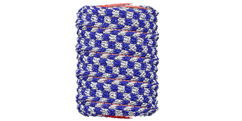 emd_11079.png emd_11079.png | 312.2 KB | ||
| マスクデータ | emd_11079_msk_1.map | 476.8 MB | マスクマップ | |
| Filedesc metadata | emd-11079.cif.gz | 6.4 KB | ||
| アーカイブディレクトリ |  http://ftp.pdbj.org/pub/emdb/structures/EMD-11079ftp://ftp.pdbj.org/pub/emdb/structures/EMD-11079 http://ftp.pdbj.org/pub/emdb/structures/EMD-11079ftp://ftp.pdbj.org/pub/emdb/structures/EMD-11079 | HTTPS FTP |
-関連構造データ

-リンク
| EMDBのページ | EMDB (EBI/PDBe) / EMDataResource |
|---|---|
| 「今月の分子」の関連する項目 |


-マップ
| ファイル | ダウンロード / ファイル: emd_11079.map.gz / 形式: CCP4 / 大きさ: 476.8 MB / タイプ: IMAGE STORED AS FLOATING POINT NUMBER (4 BYTES) | ||||||||||||||||||||||||||||||||||||||||||||||||||||||||||||
|---|---|---|---|---|---|---|---|---|---|---|---|---|---|---|---|---|---|---|---|---|---|---|---|---|---|---|---|---|---|---|---|---|---|---|---|---|---|---|---|---|---|---|---|---|---|---|---|---|---|---|---|---|---|---|---|---|---|---|---|---|---|
| 注釈 | symmetrised helical reconstruction map | ||||||||||||||||||||||||||||||||||||||||||||||||||||||||||||
| ボクセルのサイズ | X=Y=Z: 1.128 Å | ||||||||||||||||||||||||||||||||||||||||||||||||||||||||||||
| 密度 |
| ||||||||||||||||||||||||||||||||||||||||||||||||||||||||||||
| 対称性 | 空間群: 1 | ||||||||||||||||||||||||||||||||||||||||||||||||||||||||||||
| 詳細 | EMDB XML:
CCP4マップ ヘッダ情報:
| ||||||||||||||||||||||||||||||||||||||||||||||||||||||||||||
-添付データ
-マスク #1
| ファイル | emd_11079_msk_1.map | ||||||||||||
|---|---|---|---|---|---|---|---|---|---|---|---|---|---|
| 投影像・断面図 |
| ||||||||||||
| 密度ヒストグラム |
 Z
Z Y
Y X
X


- 試料の構成要素
-全体 : Helical reconstruction of influenza A virus M1 protein in complex...
| 全体 | 名称: Helical reconstruction of influenza A virus M1 protein in complex with nucleic acid |
|---|---|
| 要素 |
|
-超分子 #1: Helical reconstruction of influenza A virus M1 protein in complex...
| 超分子 | 名称: Helical reconstruction of influenza A virus M1 protein in complex with nucleic acid タイプ: complex / ID: 1 / 親要素: 0 / 含まれる分子: all / 詳細: Influenza A M1 in complex with 6.4 Kb DNA plasmid |
|---|---|
| 由来(天然) | 生物種: Influenza A virus (strain A/Puerto Rico/8/1934 H1N1) (A型インフルエンザウイルス) |
| 分子量 | 理論値: 28 KDa |
-分子 #1: Matrix protein 1
| 分子 | 名称: Matrix protein 1 / タイプ: protein_or_peptide / ID: 1 / コピー数: 1 / 光学異性体: LEVO |
|---|---|
| 由来(天然) | 生物種: Influenza A virus (strain A/Puerto Rico/8/1934 H1N1) (A型インフルエンザウイルス) |
| 分子量 | 理論値: 28.97143 KDa |
| 組換発現 | 生物種:  Escherichia coli BL21(DE3) (大腸菌) Escherichia coli BL21(DE3) (大腸菌) |
| 配列 | 文字列: MSLLTEVETY VLSIIPSGPL KAEIAQRLED VFAGKNTDLE VLMEWLKTRP ILSPLTKGIL GFVFTLTVPS ERGLQRRRFV QNALNGNGD PNNMDKAVKL YRKLKREITF HGAKEISLSY SAGALASCMG LIYNKMGAVT TEVAFGLVCA TCEQIADSQH R SHRQMVTT ...文字列: MSLLTEVETY VLSIIPSGPL KAEIAQRLED VFAGKNTDLE VLMEWLKTRP ILSPLTKGIL GFVFTLTVPS ERGLQRRRFV QNALNGNGD PNNMDKAVKL YRKLKREITF HGAKEISLSY SAGALASCMG LIYNKMGAVT TEVAFGLVCA TCEQIADSQH R SHRQMVTT TNPLIRHENR MVLASTTAKA MEQMAGSSEQ AAEAMEVASQ ARQMVQAMRT IGTHPSSSAG LKNDLLENLQ AY QKRMGVQ MQRFKLEHHH HHH UniProtKB: Matrix protein 1 |
-実験情報
-構造解析
| 手法 | クライオ電子顕微鏡法 |
|---|---|
 解析 解析 | らせん対称体再構成法 |
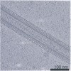
| 試料の集合状態 | helical array |
-試料調製
| 濃度 | 0.1 mg/mL | |||||||||
|---|---|---|---|---|---|---|---|---|---|---|
| 緩衝液 | pH: 10 構成要素:
| |||||||||
| グリッド | モデル: C-flat-2/2 / 材質: COPPER / メッシュ: 200 / 支持フィルム - 材質: CARBON / 支持フィルム - トポロジー: HOLEY ARRAY / 前処理 - タイプ: GLOW DISCHARGE / 前処理 - 時間: 30 sec. / 前処理 - 雰囲気: AIR | |||||||||
| 凍結 | 凍結剤: ETHANE / チャンバー内湿度: 100 % / チャンバー内温度: 298 K / 装置: FEI VITROBOT MARK IV 詳細: sample was applied 3 times each with 30s adsorption time. | |||||||||
| 詳細 | M1 at 0.1 mg/ml plasmid DNA at 0.25 mg/ml |
- 電子顕微鏡法
| 顕微鏡 | FEI TITAN KRIOS |
|---|---|
| 電子線 | 加速電圧: 300 kV / 電子線源: FIELD EMISSION GUN |
| 電子光学系 | 照射モード: FLOOD BEAM / 撮影モード: BRIGHT FIELDBright-field microscopy / Cs: 2.7 mm / 最大 デフォーカス(公称値): 3.0 µm / 最小 デフォーカス(公称値): 1.0 µm / 倍率(公称値): 105000 |
| 特殊光学系 | エネルギーフィルター - 名称: GIF Quantum LS / エネルギーフィルター - スリット幅: 20 eV |
| 試料ステージ | 試料ホルダーモデル: FEI TITAN KRIOS AUTOGRID HOLDER ホルダー冷却材: NITROGEN |
| 撮影 | フィルム・検出器のモデル: GATAN K2 SUMMIT (4k x 4k) 検出モード: COUNTING / 撮影したグリッド数: 1 / 実像数: 2347 / 平均電子線量: 47.1 e/Å2 |
| 実験機器 |  モデル: Titan Krios / 画像提供: FEI Company |
-画像解析
| Segment selection | 選択した数: 463152 / ソフトウェア - 名称: SPRING (ver. 0.86) ソフトウェア - 詳細: Used for selecting segments of similar diameters 詳細: Manual picking |
|---|---|
| 初期モデル | モデルのタイプ: NONE |
| 最終 角度割当 | タイプ: NOT APPLICABLE / ソフトウェア - 名称: RELION (ver. 3.08) |
| 最終 再構成 | 使用したクラス数: 1 想定した対称性 - らせんパラメータ - Δz: 3.08413 Å 想定した対称性 - らせんパラメータ - ΔΦ: -11.1081 ° 想定した対称性 - らせんパラメータ - 軸対称性: D1 (2回x1回 2面回転対称 )アルゴリズム: FOURIER SPACE / 解像度のタイプ: BY AUTHOR / 解像度: 3.8 Å / 解像度の算出法: FSC 0.143 CUT-OFF / ソフトウェア - 名称: RELION (ver. 3.08) / 使用した粒子像数: 17984 |