 Movie
Movie Controller

[English] 日本語
 Yorodumi
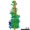
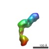
- EMDB-10522: Cryo-EM structure of Toxoplasma gondii mitochondrial ATP synthase... -
+ Open data

- Basic information
| Entry | Database: EMDB / ID: EMD-10522 | |||||||||||||||
|---|---|---|---|---|---|---|---|---|---|---|---|---|---|---|---|---|
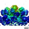
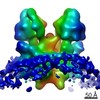
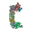
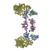
| Title | Cryo-EM structure of Toxoplasma gondii mitochondrial ATP synthase dimer, peripheral stalk map | |||||||||||||||
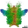
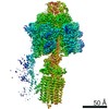
 Map data Map data | Toxoplasma gondii ATP synthase dimer, peripheral stalk full map | |||||||||||||||
 Sample Sample |
| |||||||||||||||
 Keywords Keywords |  mitochondrial / ATP synthase / peripheral stalk / OSCP / MEMBRANE PROTEIN mitochondrial / ATP synthase / peripheral stalk / OSCP / MEMBRANE PROTEIN | |||||||||||||||
| Function / homology |  Function and homology information Function and homology informationphotosynthetic electron transport in photosystem I / mitochondrial proton-transporting ATP synthase, stator stalk / mitochondrial proton-transporting ATP synthase, catalytic core / mitochondrial proton-transporting ATP synthase complex, coupling factor F(o) / photosynthetic electron transport in photosystem II / chloroplast thylakoid membrane / proton motive force-driven mitochondrial ATP synthesis / proton motive force-driven ATP synthesis / proton transmembrane transporter activity / proton-transporting ATP synthase complex, catalytic core F(1) ...photosynthetic electron transport in photosystem I / mitochondrial proton-transporting ATP synthase, stator stalk / mitochondrial proton-transporting ATP synthase, catalytic core / mitochondrial proton-transporting ATP synthase complex, coupling factor F(o) / photosynthetic electron transport in photosystem II / chloroplast thylakoid membrane / proton motive force-driven mitochondrial ATP synthesis / proton motive force-driven ATP synthesis / proton transmembrane transporter activity / proton-transporting ATP synthase complex, catalytic core F(1) / proton-transporting ATP synthase activity, rotational mechanism / ADP binding / hydrolase activity / ATP binding / plasma membraneSimilarity search - Function | |||||||||||||||
| Biological species |  Toxoplasma gondii GT1 (eukaryote) / Toxoplasma gondii (strain ATCC 50853 / GT1) (eukaryote) Toxoplasma gondii GT1 (eukaryote) / Toxoplasma gondii (strain ATCC 50853 / GT1) (eukaryote) | |||||||||||||||
| Method | single particle reconstruction / cryo EM / Resolution: 3.5 Å | |||||||||||||||
 Authors Authors | Muhleip A / Kock Flygaard R / Amunts A | |||||||||||||||
| Funding support |  Sweden, 4 items Sweden, 4 items
| |||||||||||||||
 Citation Citation | Journal: Nat Commun / Year: 2021 Title: ATP synthase hexamer assemblies shape cristae of Toxoplasma mitochondria. Authors: Alexander Mühleip / Rasmus Kock Flygaard / Jana Ovciarikova / Alice Lacombe / Paula Fernandes / Lilach Sheiner / Alexey Amunts /  Abstract: Mitochondrial ATP synthase plays a key role in inducing membrane curvature to establish cristae. In Apicomplexa causing diseases such as malaria and toxoplasmosis, an unusual cristae morphology has ...Mitochondrial ATP synthase plays a key role in inducing membrane curvature to establish cristae. In Apicomplexa causing diseases such as malaria and toxoplasmosis, an unusual cristae morphology has been observed, but its structural basis is unknown. Here, we report that the apicomplexan ATP synthase assembles into cyclic hexamers, essential to shape their distinct cristae. Cryo-EM was used to determine the structure of the hexamer, which is held together by interactions between parasite-specific subunits in the lumenal region. Overall, we identified 17 apicomplexan-specific subunits, and a minimal and nuclear-encoded subunit-a. The hexamer consists of three dimers with an extensive dimer interface that includes bound cardiolipins and the inhibitor IF. Cryo-ET and subtomogram averaging revealed that hexamers arrange into ~20-megadalton pentagonal pyramids in the curved apical membrane regions. Knockout of the linker protein ATPTG11 resulted in the loss of pentagonal pyramids with concomitant aberrantly shaped cristae. Together, this demonstrates that the unique macromolecular arrangement is critical for the maintenance of cristae morphology in Apicomplexa. | |||||||||||||||
| History |
|
- Structure visualization
| Movie |
Movie viewer |
|---|---|
| Structure viewer | EM map: SurfViewMolmilJmol/JSmol |
| Supplemental images |

- Downloads & links
-EMDB archive
| Map data | emd_10522.map.gz | 378.6 MB | EMDB map data format | |
|---|---|---|---|---|
| Header (meta data) | emd-10522-v30.xmlemd-10522.xml | 21.4 KB 21.4 KB | Display Display | EMDB header |
| FSC (resolution estimation) | emd_10522_fsc.xml | 19.9 KB | Display | FSC data file |
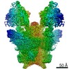
| Images |  emd_10522.png emd_10522.png | 36 KB | ||
| Masks | emd_10522_msk_1.map | 669.9 MB | Mask map | |
| Filedesc metadata | emd-10522.cif.gz | 6.9 KB | ||
| Others | emd_10522_half_map_1.map.gzemd_10522_half_map_2.map.gz | 538.9 MB 538.9 MB | ||
| Archive directory |  http://ftp.pdbj.org/pub/emdb/structures/EMD-10522ftp://ftp.pdbj.org/pub/emdb/structures/EMD-10522 http://ftp.pdbj.org/pub/emdb/structures/EMD-10522ftp://ftp.pdbj.org/pub/emdb/structures/EMD-10522 | HTTPS FTP |
-Related structure data
| Related structure data | 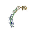 6tmiMC 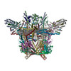 6tmgC 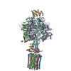 6tmhC 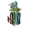 6tmjC 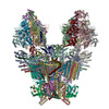 6tmkC 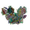 6tmlC M: atomic model generated by this map C: citing same article ( |
|---|---|
| Similar structure data |


-Links
| EMDB pages | EMDB (EBI/PDBe) / EMDataResource |
|---|---|
| Related items in Molecule of the Month |


-Map
| File | Download / File: emd_10522.map.gz / Format: CCP4 / Size: 669.9 MB / Type: IMAGE STORED AS FLOATING POINT NUMBER (4 BYTES) | ||||||||||||||||||||||||||||||||||||||||||||||||||||||||||||
|---|---|---|---|---|---|---|---|---|---|---|---|---|---|---|---|---|---|---|---|---|---|---|---|---|---|---|---|---|---|---|---|---|---|---|---|---|---|---|---|---|---|---|---|---|---|---|---|---|---|---|---|---|---|---|---|---|---|---|---|---|---|
| Annotation | Toxoplasma gondii ATP synthase dimer, peripheral stalk full map | ||||||||||||||||||||||||||||||||||||||||||||||||||||||||||||
| Voxel size | X=Y=Z: 0.83 Å | ||||||||||||||||||||||||||||||||||||||||||||||||||||||||||||
| Density |
| ||||||||||||||||||||||||||||||||||||||||||||||||||||||||||||
| Symmetry | Space group: 1 | ||||||||||||||||||||||||||||||||||||||||||||||||||||||||||||
| Details | EMDB XML:
CCP4 map header:
| ||||||||||||||||||||||||||||||||||||||||||||||||||||||||||||
-Supplemental data
-Mask #1
| File | emd_10522_msk_1.map | ||||||||||||
|---|---|---|---|---|---|---|---|---|---|---|---|---|---|
| Projections & Slices |
| ||||||||||||
| Density Histograms |
 Z
Z Y
Y X
X


-Half map: Halfmap 1
| File | emd_10522_half_map_1.map | ||||||||||||
|---|---|---|---|---|---|---|---|---|---|---|---|---|---|
| Annotation | Halfmap 1 | ||||||||||||
| Projections & Slices |
| ||||||||||||
| Density Histograms |


-Half map: Halfmap 2
| File | emd_10522_half_map_2.map | ||||||||||||
|---|---|---|---|---|---|---|---|---|---|---|---|---|---|
| Annotation | Halfmap 2 | ||||||||||||
| Projections & Slices |
| ||||||||||||
| Density Histograms |


- Sample components
-Entire : Mitochondrial ATP synthase dimer, peripheral stalk
| Entire | Name: Mitochondrial ATP synthase dimer, peripheral stalk |
|---|---|
| Components |
|
-Supramolecule #1: Mitochondrial ATP synthase dimer, peripheral stalk
| Supramolecule | Name: Mitochondrial ATP synthase dimer, peripheral stalk / type: complex / ID: 1 / Parent: 0 / Macromolecule list: all |
|---|---|
| Source (natural) | Organism: Toxoplasma gondii GT1 (eukaryote) |
| Molecular weight | Theoretical: 68 KDa |
-Macromolecule #1: ATP synthase subunit alpha
| Macromolecule | Name: ATP synthase subunit alpha / type: protein_or_peptide / ID: 1 / Number of copies: 1 / Enantiomer: LEVO |
|---|---|
| Source (natural) | Organism: Toxoplasma gondii (strain ATCC 50853 / GT1) (eukaryote) |
| Molecular weight | Theoretical: 61.189168 KDa |
| Sequence | String: MTIHSCLARR AVSVAASGAR AFASGLGARA VAVGALQSAR LLHTSSLRAA GAKISPSEMS RLLEERIAGW KTQTSTEEVG RVVSVGDGI ARLFGLEGVQ AGELVEFQNG MTGMALNLET DNVGVVIFGD DRSVLEGDSV KRTGRIVDVP IGPGLLGRVV D ALGNPIDG ...String: MTIHSCLARR AVSVAASGAR AFASGLGARA VAVGALQSAR LLHTSSLRAA GAKISPSEMS RLLEERIAGW KTQTSTEEVG RVVSVGDGI ARLFGLEGVQ AGELVEFQNG MTGMALNLET DNVGVVIFGD DRSVLEGDSV KRTGRIVDVP IGPGLLGRVV D ALGNPIDG KGPIPAKERR RVELKAPGII PRKSVHEPMM TGLKCVDALV PVGRGQRELI IGDRQTGKTA VAVDAIINQK EI NDSTDDE SKKLYCIYVA VGQKRSTVAQ IVKALEQRDA MKYTTVVAAT ASEAAPLQFL APYSGCAMGE WFRDSGRHCV IIY DDLSKQ ATAYRQMSLL LRRPPGREAY PGDVFYLHSR LLERAAKMGD KSGGGSLTAL PVIETQAGDV SAYIPTNVIS ITDG QIFLE TELFYKGIRP AINVGLSVSR VGSAAQVKAM KQVAGTMKLE LAQYREVAAF AQFGSDLDAS TRQLLTRGTA LTELL KQRQ YSPMKNSVQV CVLYCGVKGY LDPLDPKEIS RFESLFIDYI NANHQDILKT IETEKELSEK TEAKLRAAVD EFVAMN EFK KK UniProtKB: ATP synthase subunit alpha |
-Macromolecule #2: Oligomycin sensitivity conferring protein (OSCP)
| Macromolecule | Name: Oligomycin sensitivity conferring protein (OSCP) / type: protein_or_peptide / ID: 2 / Number of copies: 1 / Enantiomer: LEVO |
|---|---|
| Source (natural) | Organism: Toxoplasma gondii (strain ATCC 50853 / GT1) (eukaryote) |
| Molecular weight | Theoretical: 27.669994 KDa |
| Sequence | String: MALPLLASRR LFSSFVFRGQ PSTLSSNLSL VRIRGLHGGS LSPPSATLPR AVQLFSSRIA FSTAAAEDSG ASQTLEGRYA SALFRVAKK KNQLEKVYGD LESVRNALKD SSEFRLFVDS PAVSVQQKLD VLRQLVNRYK FDPLTGNLLT TLVENKRLPM L ARVADAFD ...String: MALPLLASRR LFSSFVFRGQ PSTLSSNLSL VRIRGLHGGS LSPPSATLPR AVQLFSSRIA FSTAAAEDSG ASQTLEGRYA SALFRVAKK KNQLEKVYGD LESVRNALKD SSEFRLFVDS PAVSVQQKLD VLRQLVNRYK FDPLTGNLLT TLVENKRLPM L ARVADAFD AMYRKEKGEV KCLVTSAKPL SAQQQKEIVA ALQNRAGTQA RLIIDYAVSP QIMGGLVVRL GEQVLDFSVA TR LDRLQSQ LLAPL UniProtKB: ATP synthase F1, delta subunit protein |
-Macromolecule #3: subunit d
| Macromolecule | Name: subunit d / type: protein_or_peptide / ID: 3 / Number of copies: 1 / Enantiomer: LEVO |
|---|---|
| Source (natural) | Organism: Toxoplasma gondii (strain ATCC 50853 / GT1) (eukaryote) |
| Molecular weight | Theoretical: 61.36225 KDa |
| Sequence | String: MQALRRGAAI PSRLLPRRDS WMSLAPFVAP NNAAAWRKLR DGAQEVQTVI ERQSTPGKPQ QIDWAKWESQ IAHKDILNCL KTFYTNQVQ ILDRALGALE TAKTPAPCEG AEKGWALFDA ALSACAKSVE KSEELLSNGA RALWVSCSNP PVWKVNTNEW L DSDQYWQA ...String: MQALRRGAAI PSRLLPRRDS WMSLAPFVAP NNAAAWRKLR DGAQEVQTVI ERQSTPGKPQ QIDWAKWESQ IAHKDILNCL KTFYTNQVQ ILDRALGALE TAKTPAPCEG AEKGWALFDA ALSACAKSVE KSEELLSNGA RALWVSCSNP PVWKVNTNEW L DSDQYWQA FVEKHHFYSQ YQPGVVDPEA PQEVEAFKQA WHSRMGKFND RSDTPMLYAY MNELPSWEYY DLHRSAFLEH MT YFLVRTG GDFRFFPEMP PWQWLAHMEN LRFKLLSVAQ SRRSQLQLAN LERERALDFL PVDVEHHGEE YTQKFLQYET ELF QACAAR LMGHFMFLCD PFIPVQSAEA LSAVTRVDNG KGKLFSLGDD VNALFYLPEQ QRRDVERPTQ AVQTLLGHLE ATGR PFNPC YSELLHVHAE VLEERGEHWL TAPGECVSQA FLRRLRTDDP AYEVYCSYFK EMYERFAGAK EVSMEDGRKR LATIE KNAQ EEAAAYGLAL KTMGSAELAH KAREGAAKLE QLRKAQEKAA GKSAQTVQEN KM UniProtKB: Uncharacterized protein |
-Macromolecule #4: ATPTG12
| Macromolecule | Name: ATPTG12 / type: protein_or_peptide / ID: 4 / Number of copies: 1 / Enantiomer: LEVO |
|---|---|
| Source (natural) | Organism: Toxoplasma gondii (strain ATCC 50853 / GT1) (eukaryote) |
| Molecular weight | Theoretical: 15.400639 KDa |
| Sequence | String: MLNFIPKRCP SVSLLFGKRP VQRIEVGQAR HQLEIPVETI EKIYEGVDSR LEYHNKDYNA MKWKDFMKLK LDAYHLLEAS QSETAAKSA LSDLNWFSDL ADIYSGQQTM AEMDVALKAQ GEQKLSYPIQ GKNIK UniProtKB: Uncharacterized protein |
-Macromolecule #5: subunit b
| Macromolecule | Name: subunit b / type: protein_or_peptide / ID: 5 / Number of copies: 1 / Enantiomer: LEVO |
|---|---|
| Source (natural) | Organism: Toxoplasma gondii (strain ATCC 50853 / GT1) (eukaryote) |
| Molecular weight | Theoretical: 64.811602 KDa |
| Sequence | String: MNFSSSARWL AVRQSQTLGH TTRATVAAGR RVLAHSPAAT EFTSFQSLHI GGDVCKLPLA VALGAAPSAL GYGSAKHNQQ RQYATLGSG WSFSKVQYTK YRITKPWTTD TTFDDIILSQ PSKEDFAKFT KEAPLFLRFL KLVTDVEGRQ EAFIQFAKRC E NGLTVEKD ...String: MNFSSSARWL AVRQSQTLGH TTRATVAAGR RVLAHSPAAT EFTSFQSLHI GGDVCKLPLA VALGAAPSAL GYGSAKHNQQ RQYATLGSG WSFSKVQYTK YRITKPWTTD TTFDDIILSQ PSKEDFAKFT KEAPLFLRFL KLVTDVEGRQ EAFIQFAKRC E NGLTVEKD VYVTKKELVD CLWKNGYTDT EINAFEIAFP ADYKFHYPEL AVLFDLTEED CYKYCIRQRA ATPEELVELK YT KPKNLVS SYGLCFLGVW FGLSNTVLSN AWFYSKTFPF GAVFYMLGSY FYRDIREKLW KEEKSLIHTA QENKNMGEES VYK QMKKYA TDTKCLDYLS TFRTEVEDQI ANYKVALVSQ MRRQLTERLV EKLNGIQQAE KLIQGSLQDV MIREIVSSFK DLYK SRPEL HDAAMQSAIQ GLSGSDGAMD PVGAHFKASL QELAKVNLST ATADPMGTVV QRVAAVFQKR EKEFLDTFTV KATEA QEIK TIVDKCHKGN TFDFHALSDE ELRRLEQLYS TVNNRVGFET IHENSIKPVA PLSENSKGFV EFVNTQLEIT KAKLRN ARL TAFAHAFV UniProtKB: Uncharacterized protein |
-Experimental details
-Structure determination
| Method | cryo EM |
|---|---|
 Processing Processing | single particle reconstruction |
| Aggregation state | particle |
-Sample preparation
| Concentration | 5 mg/mL |
|---|---|
| Buffer | pH: 7.5 |
| Vitrification | Cryogen name: ETHANE / Chamber humidity: 100 % / Instrument: FEI VITROBOT MARK IV / Details: 3 seconds blot.. |
- Electron microscopy
| Microscope | FEI TITAN KRIOS |
|---|---|
| Electron beam | Acceleration voltage: 300 kV / Electron source: FIELD EMISSION GUN |
| Electron optics | Illumination mode: FLOOD BEAM / Imaging mode: BRIGHT FIELDBright-field microscopy / Cs: 2.7 mm / Nominal magnification: 165000 |
| Specialist optics | Energy filter - Name: GIF Quantum LS / Energy filter - Slit width: 20 eV |
| Sample stage | Specimen holder model: FEI TITAN KRIOS AUTOGRID HOLDER / Cooling holder cryogen: NITROGEN |
| Image recording | Film or detector model: GATAN K2 QUANTUM (4k x 4k) / Detector mode: COUNTING / Number real images: 4860 / Average electron dose: 30.0 e/Å2 |
| Experimental equipment |  Model: Titan Krios / Image courtesy: FEI Company |
-Image processing
| Startup model | Type of model: OTHER |
|---|---|
| Initial angle assignment | Type: MAXIMUM LIKELIHOOD / Software - Name: RELION (ver. 3.0) |
| Final angle assignment | Type: MAXIMUM LIKELIHOOD / Software - Name: RELION (ver. 3.0) |
| Final reconstruction | Applied symmetry - Point group: C1 (asymmetric) / Algorithm: FOURIER SPACE / Resolution.type: BY AUTHOR / Resolution: 3.5 Å / Resolution method: FSC 0.143 CUT-OFF / Software - Name: RELION (ver. 3.0) / Number images used: 203010 |
| FSC plot (resolution estimation) |  |
-Atomic model buiding 1
| Refinement | Space: REAL |
|---|---|
| Output model | PDB-6tmi: |
