 Movie
Movie Controller

[English] 日本語
 Yorodumi
- PDB-7m05: CryoEM structure of PRMT5 bound to covalent PBM-site inhibitor BR... -
+ Open data

- Basic information
| Entry | Database: PDB / ID: 7m05 | ||||||
|---|---|---|---|---|---|---|---|
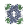
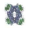
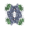
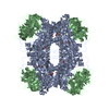
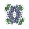
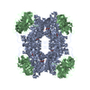
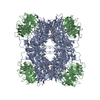
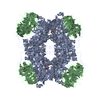
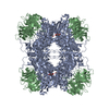
| Title | CryoEM structure of PRMT5 bound to covalent PBM-site inhibitor BRD-6988 | ||||||
 Components Components |
| ||||||
 Keywords Keywords | TRANSFERASE/INHIBITOR /  methyltransferase / splicing / epigenetic / TRANSFERASE-INHIBITOR complex methyltransferase / splicing / epigenetic / TRANSFERASE-INHIBITOR complex | ||||||
| Function / homology |  Function and homology information Function and homology informationpositive regulation of adenylate cyclase-inhibiting dopamine receptor signaling pathway / peptidyl-arginine N-methylation / oocyte axis specification / type II protein arginine methyltransferase / protein-arginine omega-N symmetric methyltransferase activity / peptidyl-arginine methylation / Golgi ribbon formation / negative regulation of epithelial cell proliferation involved in prostate gland development / histone H4R3 methyltransferase activity / secretory columnal luminar epithelial cell differentiation involved in prostate glandular acinus development ...positive regulation of adenylate cyclase-inhibiting dopamine receptor signaling pathway / peptidyl-arginine N-methylation / oocyte axis specification / type II protein arginine methyltransferase / protein-arginine omega-N symmetric methyltransferase activity / peptidyl-arginine methylation / Golgi ribbon formation / negative regulation of epithelial cell proliferation involved in prostate gland development / histone H4R3 methyltransferase activity / secretory columnal luminar epithelial cell differentiation involved in prostate glandular acinus development / epithelial cell proliferation involved in prostate gland development / histone arginine N-methyltransferase activity / methylosome / protein-arginine N-methyltransferase activity / positive regulation of mRNA splicing, via spliceosome / methyl-CpG binding / : / endothelial cell activation / histone H3 methyltransferase activity / Cul4B-RING E3 ubiquitin ligase complex / histone methyltransferase complex / regulation of mitotic nuclear division / positive regulation of oligodendrocyte differentiation / histone methyltransferase activity / E-box binding / negative regulation of cell differentiation / ubiquitin-like ligase-substrate adaptor activity / spliceosomal snRNP assembly / ribonucleoprotein complex binding / regulation of ERK1 and ERK2 cascade / nuclear receptor coactivator activity / regulation of signal transduction by p53 class mediator / liver regeneration / methyltransferase activity / DNA-templated transcription termination / circadian regulation of gene expression / Regulation of TP53 Activity through Methylation / RMTs methylate histone arginines / protein polyubiquitination / transcription corepressor activity / p53 binding / snRNP Assembly / ubiquitin-dependent protein catabolic process / chromatin remodeling / protein heterodimerization activity / positive regulation of cell population proliferation / chromatin / regulation of DNA-templated transcription / regulation of transcription by RNA polymerase II / Golgi apparatus / nucleoplasm / identical protein binding / nucleus / cytosol / cytoplasmSimilarity search - Function | ||||||
| Biological species |  Homo sapiens (human) Homo sapiens (human) | ||||||
| Method | ELECTRON MICROSCOPY / single particle reconstruction / cryo EM / Resolution: 2.39 Å | ||||||
 Authors Authors | McMillan, B.J. / McKinney, D.C. / Timm, D.E. | ||||||
 Citation Citation | Journal: J Med Chem / Year: 2021 Title: Discovery of a First-in-Class Inhibitor of the PRMT5-Substrate Adaptor Interaction. Authors: David C McKinney / Brian J McMillan / Matthew J Ranaghan / Jamie A Moroco / Merissa Brousseau / Zachary Mullin-Bernstein / Meghan O'Keefe / Patrick McCarren / Michael F Mesleh / Kathleen M ...Authors: David C McKinney / Brian J McMillan / Matthew J Ranaghan / Jamie A Moroco / Merissa Brousseau / Zachary Mullin-Bernstein / Meghan O'Keefe / Patrick McCarren / Michael F Mesleh / Kathleen M Mulvaney / Foxy Robinson / Ritu Singh / Besnik Bajrami / Florence F Wagner / Robert Hilgraf / Martin J Drysdale / Arthur J Campbell / Adam Skepner / David E Timm / Dale Porter / Virendar K Kaushik / William R Sellers / Alessandra Ianari /  Abstract: PRMT5 and its substrate adaptor proteins (SAPs), pICln and Riok1, are synthetic lethal dependencies in MTAP-deleted cancer cells. SAPs share a conserved PRMT5 binding motif (PBM) which mediates ...PRMT5 and its substrate adaptor proteins (SAPs), pICln and Riok1, are synthetic lethal dependencies in MTAP-deleted cancer cells. SAPs share a conserved PRMT5 binding motif (PBM) which mediates binding to a surface of PRMT5 distal to the catalytic site. This interaction is required for methylation of several PRMT5 substrates, including histone and spliceosome complexes. We screened for small molecule inhibitors of the PRMT5-PBM interaction and validated a compound series which binds to the PRMT5-PBM interface and directly inhibits binding of SAPs. Mode of action studies revealed the formation of a covalent bond between a halogenated pyridazinone group and cysteine 278 of PRMT5. Optimization of the starting hit produced a lead compound, BRD0639, which engages the target in cells, disrupts PRMT5-RIOK1 complexes, and reduces substrate methylation. BRD0639 is a first-in-class PBM-competitive inhibitor that can support studies of PBM-dependent PRMT5 activities and the development of novel PRMT5 inhibitors that selectively target these functions. #1: Journal: bioRxiv / Year: 2020Title: Discovery of a first-in-class inhibitor of the PRMT5-substrate adaptor interaction Authors: Mulvaney, K.M. / McMillan, B.J. / Sellers, W.R. | ||||||
| History |
|
- Structure visualization
| Movie |
Movie viewer |
|---|---|
| Structure viewer | Molecule: MolmilJmol/JSmol |
- Downloads & links
-Download
| PDBx/mmCIF format | 7m05.cif.gz | 1.1 MB | Display | PDBx/mmCIF format |
|---|---|---|---|---|
| PDB format | pdb7m05.ent.gz | 1002.7 KB | Display | PDB format |
| PDBx/mmJSON format | 7m05.json.gz | Tree view | PDBx/mmJSON format | |
| Others |  Other downloads Other downloads |
-Validation report
| Arichive directory | https://data.pdbj.org/pub/pdb/validation_reports/m0/7m05ftp://data.pdbj.org/pub/pdb/validation_reports/m0/7m05 | HTTPS FTP |
|---|
-Related structure data
| Related structure data | 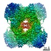 23609MC  6v0pC M: map data used to model this data C: citing same article ( |
|---|---|
| Similar structure data |
-Links
 PDBj
PDBj
- Assembly
| Deposited unit | 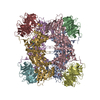
|
|---|---|
| 1 |
|
-Components
| #1: Protein | Mass: 72766.664 Da / Num. of mol.: 4 Source method: isolated from a genetically manipulated source Source: (gene. exp.) Homo sapiens (human) / Gene: PRMT5, HRMT1L5, IBP72, JBP1, SKB1 / Production host:   Spodoptera frugiperda (fall armyworm) Spodoptera frugiperda (fall armyworm)References: UniProt: O14744, type II protein arginine methyltransferase #2: Protein | WD repeat-containing protein 77 / MEP-50 / Androgen receptor cofactor p44 / WD repeat-containing protein 77 / p44/Mep50Mass: 36723.164 Da / Num. of mol.: 4 Source method: isolated from a genetically manipulated source Source: (gene. exp.) Homo sapiens (human) / Gene: WDR77, MEP50, WD45, HKMT1069, Nbla10071 / Production host: Spodoptera frugiperda (fall armyworm) / References: UniProt: Q9BQA1#3: Chemical | ChemComp-YJG /   Mass: 461.922 Da / Num. of mol.: 4 / Source method: obtained synthetically / Formula: C20H20ClN5O4S / Feature type: SUBJECT OF INVESTIGATION Mass: 461.922 Da / Num. of mol.: 4 / Source method: obtained synthetically / Formula: C20H20ClN5O4S / Feature type: SUBJECT OF INVESTIGATIONHas ligand of interest | Y | |
|---|
-Experimental details
-Experiment
| Experiment | Method: ELECTRON MICROSCOPY |
|---|---|
| EM experiment | Aggregation state: PARTICLE / 3D reconstruction method: single particle reconstruction |
- Sample preparation
| Component | Name: Hetero-octamer complex of PRMT5 and WDR77 / Type: COMPLEX / Entity ID: #1-#2 / Source: RECOMBINANT | |||||||||||||||||||||||||
|---|---|---|---|---|---|---|---|---|---|---|---|---|---|---|---|---|---|---|---|---|---|---|---|---|---|---|
| Molecular weight | Value: 0.437 MDa / Experimental value: NO | |||||||||||||||||||||||||
| Source (natural) | Organism: Homo sapiens (human) | |||||||||||||||||||||||||
| Source (recombinant) | Organism: Spodoptera frugiperda (fall armyworm) | |||||||||||||||||||||||||
| Buffer solution | pH: 7.4 | |||||||||||||||||||||||||
| Buffer component |
| |||||||||||||||||||||||||
| Specimen | Conc.: 1.42 mg/ml / Embedding applied: NO / Shadowing applied: NO / Staining applied: NO / Vitrification applied: YES | |||||||||||||||||||||||||
| Specimen support | Grid material: GOLD / Grid mesh size: 300 divisions/in. / Grid type: UltrAuFoil | |||||||||||||||||||||||||
| Vitrification | Instrument: FEI VITROBOT MARK IV / Cryogen name: ETHANE / Humidity: 100 % / Chamber temperature: 277.15 K |
- Electron microscopy imaging
| Experimental equipment |  Model: Titan Krios / Image courtesy: FEI Company |
|---|---|
| Microscopy | Model: FEI TITAN KRIOS |
| Electron gun | Electron source: FIELD EMISSION GUN / Accelerating voltage: 300 kV / Illumination mode: FLOOD BEAM |
| Electron lens | Mode: BRIGHT FIELDBright-field microscopy / Nominal magnification: 130000 X / Calibrated magnification: 46296 X / Nominal defocus max: 2100 nm / Nominal defocus min: 1400 nm / Cs: 2.7 mm / Alignment procedure: COMA FREE |
| Specimen holder | Cryogen: NITROGEN / Specimen holder model: FEI TITAN KRIOS AUTOGRID HOLDER |
| Image recording | Average exposure time: 8 sec. / Electron dose: 62.4 e/Å2 / Detector mode: COUNTING / Film or detector model: GATAN K2 SUMMIT (4k x 4k) / Num. of grids imaged: 1 / Num. of real images: 2169 |
| EM imaging optics | Energyfilter name: GIF Quantum LS / Energyfilter slit width: 30 eV |
| Image scans | Sampling size: 5 µm / Width: 3838 / Height: 3710 / Movie frames/image: 40 / Used frames/image: 4-40 |
- Processing
| Software | Name: PHENIX / Version: 1.16_3549: / Classification: refinement | ||||||||||||||||||||||||||||||||||||||||
|---|---|---|---|---|---|---|---|---|---|---|---|---|---|---|---|---|---|---|---|---|---|---|---|---|---|---|---|---|---|---|---|---|---|---|---|---|---|---|---|---|---|
| EM software |
| ||||||||||||||||||||||||||||||||||||||||
| CTF correction | Type: PHASE FLIPPING AND AMPLITUDE CORRECTION | ||||||||||||||||||||||||||||||||||||||||
| Particle selection | Num. of particles selected: 956646 | ||||||||||||||||||||||||||||||||||||||||
| Symmetry | Point symmetry: D2 (2x2 fold dihedral) | ||||||||||||||||||||||||||||||||||||||||
| 3D reconstruction | Resolution: 2.39 Å / Resolution method: FSC 0.143 CUT-OFF / Num. of particles: 443624 / Algorithm: FOURIER SPACE / Num. of class averages: 22 / Symmetry type: POINT | ||||||||||||||||||||||||||||||||||||||||
| Atomic model building | Protocol: RIGID BODY FIT / Space: REAL | ||||||||||||||||||||||||||||||||||||||||
| Atomic model building | PDB-ID: 6V0P | ||||||||||||||||||||||||||||||||||||||||
| Refine LS restraints |
|
