[English] 日本語
Yorodumi
- PDB-6ynx: Cryo-EM structure of Tetrahymena thermophila mitochondrial ATP sy... -

+
Open data


ID or keywords:

Loading...

-
Basic information

Entry
Database: PDB / ID: 6ynx
TitleCryo-EM structure of Tetrahymena thermophila mitochondrial ATP synthase - Fo-subcomplex
Components
  • ATPTT1
  • ATPTT10
  • ATPTT11
  • ATPTT12
  • ATPTT13
  • ATPTT2
  • ATPTT3
  • ATPTT4
  • ATPTT5
  • ATPTT6
  • ATPTT7
  • ATPTT8
  • ATPTT9
  • Inhibitor of F1 (IF1)
  • subunit 8
  • subunit a
  • subunit b
  • subunit d
  • subunit f
  • subunit i/j
  • subunit k
KeywordsMEMBRANE PROTEIN / mitochondria / ATP synthase / Fo-subcomplex / IF1 dimer
Function / homology
Function and homology information


mitochondrial proton-transporting ATP synthase complex, coupling factor F(o) / proton motive force-driven ATP synthesis / proton transmembrane transporter activity / oxidoreductase activity / hydrolase activity / mitochondrion / membrane
Similarity search - Function
Sulphide quinone-reductase / ATP synthase, F0 complex, subunit D, mitochondrial / ATP synthase D chain, mitochondrial (ATP5H) / ATP synthase, F0 complex, subunit D superfamily, mitochondrial / FAD/NAD(P)-binding domain / Pyridine nucleotide-disulphide oxidoreductase / alpha/beta hydrolase fold / Alpha/beta hydrolase fold-1 / ACP-like superfamily / Carrier protein (CP) domain profile. ...Sulphide quinone-reductase / ATP synthase, F0 complex, subunit D, mitochondrial / ATP synthase D chain, mitochondrial (ATP5H) / ATP synthase, F0 complex, subunit D superfamily, mitochondrial / FAD/NAD(P)-binding domain / Pyridine nucleotide-disulphide oxidoreductase / alpha/beta hydrolase fold / Alpha/beta hydrolase fold-1 / ACP-like superfamily / Carrier protein (CP) domain profile. / Phosphopantetheine binding ACP domain / FAD/NAD(P)-binding domain superfamily / Alpha/Beta hydrolase fold
Similarity search - Domain/homology
ADENOSINE-5'-TRIPHOSPHATE / CARDIOLIPIN / NICOTINAMIDE-ADENINE-DINUCLEOTIDE / 1,2-DIACYL-SN-GLYCERO-3-PHOSPHOCHOLINE / 1,2-dioleoyl-sn-glycero-3-phosphoethanolamine / PHOSPHATE ION / Ubiquinone-8 / Transmembrane protein, putative / Uncharacterized protein / Oxidoreductase, putative ...ADENOSINE-5'-TRIPHOSPHATE / CARDIOLIPIN / NICOTINAMIDE-ADENINE-DINUCLEOTIDE / 1,2-DIACYL-SN-GLYCERO-3-PHOSPHOCHOLINE / 1,2-dioleoyl-sn-glycero-3-phosphoethanolamine / PHOSPHATE ION / Ubiquinone-8 / Transmembrane protein, putative / Uncharacterized protein / Oxidoreductase, putative / Transmembrane protein / Transmembrane protein, putative / Transmembrane protein / Alpha/beta hydrolase / Uncharacterized protein / Transmembrane protein, putative / Transmembrane protein / Transmembrane protein / Uncharacterized protein / Transmembrane protein, putative / Uncharacterized protein / Uncharacterized protein / Acyl carrier protein, putative / Uncharacterized protein / Uncharacterized protein / Transmembrane protein / Ymf56 / Ymf66
Similarity search - Component
Biological speciesTetrahymena thermophila (eukaryote)
MethodELECTRON MICROSCOPY / single particle reconstruction / cryo EM / Resolution: 2.5 Å
AuthorsKock Flygaard, R. / Muhleip, A. / Amunts, A.
Funding support Sweden, 3items
OrganizationGrant numberCountry
Swedish Research CouncilNT_2015-04107 Sweden
European Research Council (ERC)ERC-2018-StG-805230 Sweden
Knut and Alice Wallenberg Foundation2018.0080 Sweden
CitationJournal: Nat Commun / Year: 2020
Title: Type III ATP synthase is a symmetry-deviated dimer that induces membrane curvature through tetramerization.
Authors: Rasmus Kock Flygaard / Alexander Mühleip / Victor Tobiasson / Alexey Amunts /
Abstract: Mitochondrial ATP synthases form functional homodimers to induce cristae curvature that is a universal property of mitochondria. To expand on the understanding of this fundamental phenomenon, we ...Mitochondrial ATP synthases form functional homodimers to induce cristae curvature that is a universal property of mitochondria. To expand on the understanding of this fundamental phenomenon, we characterized the unique type III mitochondrial ATP synthase in its dimeric and tetrameric form. The cryo-EM structure of a ciliate ATP synthase dimer reveals an unusual U-shaped assembly of 81 proteins, including a substoichiometrically bound ATPTT2, 40 lipids, and co-factors NAD and CoQ. A single copy of subunit ATPTT2 functions as a membrane anchor for the dimeric inhibitor IF. Type III specific linker proteins stably tie the ATP synthase monomers in parallel to each other. The intricate dimer architecture is scaffolded by an extended subunit-a that provides a template for both intra- and inter-dimer interactions. The latter results in the formation of tetramer assemblies, the membrane part of which we determined to 3.1 Å resolution. The structure of the type III ATP synthase tetramer and its associated lipids suggests that it is the intact unit propagating the membrane curvature.
History
DepositionApr 14, 2020Deposition site: PDBE / Processing site: PDBE
Revision 1.0Sep 30, 2020Provider: repository / Type: Initial release
Revision 1.1Nov 11, 2020Group: Database references / Category: citation / citation_author
Item: _citation.country / _citation.journal_abbrev ..._citation.country / _citation.journal_abbrev / _citation.journal_id_CSD / _citation.journal_id_ISSN / _citation.journal_volume / _citation.page_first / _citation.page_last / _citation.pdbx_database_id_DOI / _citation.pdbx_database_id_PubMed / _citation.title / _citation.year

-
Structure visualization

Movie
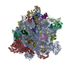
  • Deposited structure unit
  • Imaged by Jmol
  • Download
  • Simplified surface model + fitted atomic model
  • EMDB-10859
  • Imaged by Jmol
  • Download
  • Superimposition on EM map
  • EMDB-10859
  • Imaged by UCSF Chimera
  • Download
Movie viewer
Structure viewerMolecule:
MolmilJmol/JSmol

Downloads & links

-
Assembly

Deposited unit
a: subunit a
b: subunit b
d: subunit d
f: subunit f
i: subunit i/j
k: subunit k
c: subunit 8
g: ATPTT3
h: ATPTT4
j: ATPTT5
l: ATPTT6
m: ATPTT7
n: ATPTT8
o: ATPTT9
p: ATPTT10
q: ATPTT11
r: ATPTT12
s: ATPTT13
e: ATPTT1
A: subunit a
B: subunit b
D: subunit d
F: subunit f
I: subunit i/j
K: subunit k
C: subunit 8
G: ATPTT3
H: ATPTT4
J: ATPTT5
L: ATPTT6
M: ATPTT7
N: ATPTT8
O: ATPTT9
P: ATPTT10
Q: ATPTT11
R: ATPTT12
S: ATPTT13
E: ATPTT1
i1: Inhibitor of F1 (IF1)
i2: Inhibitor of F1 (IF1)
t: ATPTT2
hetero molecules


Theoretical massNumber of molelcules
Total (without water)1,183,26991
Polymers1,127,57641
Non-polymers55,69250
Water0
1


  • Idetical with deposited unit
  • defined by author
  • Evidence: microscopy
TypeNameSymmetry operationNumber
identity operation1_5551

-
Components

+
Protein , 21 types, 41 molecules aAbBdDfFiIkKcCgGhHjJlLmMnNoOpP...

#1: Protein subunit a


Mass: 52561.461 Da / Num. of mol.: 2 / Source method: isolated from a natural source / Source: (natural) Tetrahymena thermophila (eukaryote) / References: UniProt: Q951C1
#2: Protein subunit b


Mass: 43910.102 Da / Num. of mol.: 2 / Source method: isolated from a natural source / Source: (natural) Tetrahymena thermophila (eukaryote) / References: UniProt: I7MJ84
#3: Protein subunit d


Mass: 26747.951 Da / Num. of mol.: 2 / Source method: isolated from a natural source / Source: (natural) Tetrahymena thermophila (eukaryote) / References: UniProt: Q239R1
#4: Protein subunit f


Mass: 24265.398 Da / Num. of mol.: 2 / Source method: isolated from a natural source / Source: (natural) Tetrahymena thermophila (eukaryote) / References: UniProt: Q24I07
#5: Protein subunit i/j


Mass: 24395.543 Da / Num. of mol.: 2 / Source method: isolated from a natural source / Source: (natural) Tetrahymena thermophila (eukaryote) / References: UniProt: I7LZW2
#6: Protein subunit k


Mass: 20963.871 Da / Num. of mol.: 2 / Source method: isolated from a natural source / Source: (natural) Tetrahymena thermophila (eukaryote) / References: UniProt: I7LSX6
#7: Protein subunit 8


Mass: 12299.238 Da / Num. of mol.: 2 / Source method: isolated from a natural source / Source: (natural) Tetrahymena thermophila (eukaryote) / References: UniProt: Q950Y8
#8: Protein ATPTT3


Mass: 34719.633 Da / Num. of mol.: 2 / Source method: isolated from a natural source / Source: (natural) Tetrahymena thermophila (eukaryote) / References: UniProt: I7M8Q3
#9: Protein ATPTT4


Mass: 32484.051 Da / Num. of mol.: 2 / Source method: isolated from a natural source / Source: (natural) Tetrahymena thermophila (eukaryote) / References: UniProt: I7MCZ0
#10: Protein ATPTT5


Mass: 31674.639 Da / Num. of mol.: 2 / Source method: isolated from a natural source / Source: (natural) Tetrahymena thermophila (eukaryote) / References: UniProt: Q228N4
#11: Protein ATPTT6


Mass: 29354.383 Da / Num. of mol.: 2 / Source method: isolated from a natural source / Source: (natural) Tetrahymena thermophila (eukaryote) / References: UniProt: I7MCQ6
#12: Protein ATPTT7


Mass: 26355.104 Da / Num. of mol.: 2 / Source method: isolated from a natural source / Source: (natural) Tetrahymena thermophila (eukaryote) / References: UniProt: I7M980
#13: Protein ATPTT8


Mass: 21782.971 Da / Num. of mol.: 2 / Source method: isolated from a natural source / Source: (natural) Tetrahymena thermophila (eukaryote) / References: UniProt: I7LVK6
#14: Protein ATPTT9


Mass: 18199.361 Da / Num. of mol.: 2 / Source method: isolated from a natural source / Source: (natural) Tetrahymena thermophila (eukaryote) / References: UniProt: Q24HK1
#15: Protein ATPTT10


Mass: 17425.703 Da / Num. of mol.: 2 / Source method: isolated from a natural source / Source: (natural) Tetrahymena thermophila (eukaryote) / References: UniProt: I7LZE5
#16: Protein ATPTT11


Mass: 17410.811 Da / Num. of mol.: 2 / Source method: isolated from a natural source / Source: (natural) Tetrahymena thermophila (eukaryote) / References: UniProt: Q22DV8
#17: Protein ATPTT12


Mass: 17343.820 Da / Num. of mol.: 2 / Source method: isolated from a natural source / Source: (natural) Tetrahymena thermophila (eukaryote) / References: UniProt: I7M0G0
#18: Protein ATPTT13


Mass: 16816.250 Da / Num. of mol.: 2 / Source method: isolated from a natural source / Source: (natural) Tetrahymena thermophila (eukaryote) / References: UniProt: I7MLU7
#19: Protein ATPTT1


Mass: 54917.816 Da / Num. of mol.: 2 / Source method: isolated from a natural source / Source: (natural) Tetrahymena thermophila (eukaryote) / References: UniProt: I7LVQ8
#20: Protein Inhibitor of F1 (IF1)


Mass: 12987.691 Da / Num. of mol.: 2 / Source method: isolated from a natural source / Source: (natural) Tetrahymena thermophila (eukaryote) / References: UniProt: I7M7C0
#21: Protein ATPTT2


Mass: 54344.680 Da / Num. of mol.: 1 / Source method: isolated from a natural source / Source: (natural) Tetrahymena thermophila (eukaryote) / References: UniProt: I7M7B9

-
Non-polymers , 8 types, 50 molecules

#22: Chemical...
ChemComp-CDL / CARDIOLIPIN / DIPHOSPHATIDYL GLYCEROL / BIS-(1,2-DIACYL-SN-GLYCERO-3-PHOSPHO)-1',3'-SN-GLYCEROL / Cardiolipin


Mass: 1464.043 Da / Num. of mol.: 30 / Source method: obtained synthetically / Formula: C81H156O17P2 / Comment: phospholipid*YM
#23: Chemical
ChemComp-PC1 / 1,2-DIACYL-SN-GLYCERO-3-PHOSPHOCHOLINE / 3-SN-PHOSPHATIDYLCHOLINE / Phosphatidylcholine


Mass: 790.145 Da / Num. of mol.: 6 / Source method: obtained synthetically / Formula: C44H88NO8P / Comment: phospholipid*YM
#24: Chemical ChemComp-PO4 / PHOSPHATE ION / Phosphate


Mass: 94.971 Da / Num. of mol.: 2 / Source method: obtained synthetically / Formula: PO4
#25: Chemical ChemComp-UQ8 / Ubiquinone-8 / 2,3-dimethoxy-5-methyl-6-[(6E,10E,14E,18E,22E,26E)-3,7,11,15,19,23,27,31-octamethyldotriaconta-2,6,10,14,18,22,26,30-oc taen-1-yl]cyclohexa-2,5-diene-1,4-dione


Mass: 727.109 Da / Num. of mol.: 2 / Source method: obtained synthetically / Formula: C49H74O4
#26: Chemical ChemComp-ATP / ADENOSINE-5'-TRIPHOSPHATE / Adenosine triphosphate


Mass: 507.181 Da / Num. of mol.: 2 / Source method: obtained synthetically / Formula: C10H16N5O13P3 / Comment: ATP, energy-carrying molecule*YM
#27: Chemical ChemComp-MG / MAGNESIUM ION


Mass: 24.305 Da / Num. of mol.: 2 / Source method: obtained synthetically / Formula: Mg
#28: Chemical
ChemComp-PEE / 1,2-Dioleoyl-sn-glycero-3-phosphoethanolamine / DOPE / Discrete optimized protein energy


Mass: 749.073 Da / Num. of mol.: 4 / Source method: obtained synthetically / Formula: C41H83NO8P / Comment: DOPE, phospholipid*YM
#29: Chemical ChemComp-NAD / NICOTINAMIDE-ADENINE-DINUCLEOTIDE / Nicotinamide adenine dinucleotide


Mass: 663.425 Da / Num. of mol.: 2 / Source method: obtained synthetically / Formula: C21H27N7O14P2 / Comment: NAD*YM

-
Details

Has ligand of interestN

-
Experimental details

-
Experiment

ExperimentMethod: ELECTRON MICROSCOPY
EM experimentAggregation state: PARTICLE / 3D reconstruction method: single particle reconstruction

-
Sample preparation

ComponentName: Mitochondrial ATP synthase, Fo-subcomplex / Type: COMPLEX / Entity ID: #1-#21 / Source: NATURAL
Molecular weightExperimental value: NO
Source (natural)Organism: Tetrahymena thermophila (eukaryote)
Buffer solutionpH: 7.5
SpecimenConc.: 0.75 mg/ml / Embedding applied: NO / Shadowing applied: NO / Staining applied: NO / Vitrification applied: YES
Specimen supportGrid material: COPPER / Grid mesh size: 300 divisions/in. / Grid type: Quantifoil R2/2
VitrificationInstrument: FEI VITROBOT MARK IV / Cryogen name: ETHANE

-
Electron microscopy imaging

Experimental equipment
Model: Titan Krios / Image courtesy: FEI Company
MicroscopyModel: FEI TITAN KRIOS
Electron gunElectron source: FIELD EMISSION GUN / Accelerating voltage: 300 kV / Illumination mode: FLOOD BEAM
Electron lensMode: BRIGHT FIELDBright-field microscopy / Nominal magnification: 165000 X
Image recordingElectron dose: 30.9 e/Å2 / Detector mode: COUNTING / Film or detector model: GATAN K2 QUANTUM (4k x 4k)
EM imaging opticsEnergyfilter name: GIF Quantum LS / Energyfilter slit width: 20 eV
Image scansMovie frames/image: 20

-
Processing

EM software
IDNameVersionCategoryDetails
1RELION3.1particle selectionbeta
2EPUimage acquisition
4CTFFIND4.1.13CTF correction
10RELION3.1initial Euler assignmentbeta
11RELION3.1final Euler assignmentbeta
13RELION3.13D reconstructionbeta
CTF correctionType: PHASE FLIPPING AND AMPLITUDE CORRECTION
SymmetryPoint symmetry: C1 (asymmetric)
3D reconstructionResolution: 2.5 Å / Resolution method: FSC 0.143 CUT-OFF / Num. of particles: 61157 / Symmetry type: POINT

+
About Yorodumi

-
News

-
Feb 9, 2022. New format data for meta-information of EMDB entries

New format data for meta-information of EMDB entries

  • Version 3 of the EMDB header file is now the official format.
  • The previous official version 1.9 will be removed from the archive.

Related info.:EMDB header

External links:wwPDB to switch to version 3 of the EMDB data model

-
Aug 12, 2020. Covid-19 info

Covid-19 info

URL: https://pdbj.org/emnavi/covid19.php

New page: Covid-19 featured information page in EM Navigator.

Related info.:Covid-19 info / Mar 5, 2020. Novel coronavirus structure data

+
Mar 5, 2020. Novel coronavirus structure data

Novel coronavirus structure data

Related info.:Yorodumi Speices / Aug 12, 2020. Covid-19 info

External links:COVID-19 featured content - PDBj / Molecule of the Month (242):Coronavirus Proteases

+
Jan 31, 2019. EMDB accession codes are about to change! (news from PDBe EMDB page)

EMDB accession codes are about to change! (news from PDBe EMDB page)

  • The allocation of 4 digits for EMDB accession codes will soon come to an end. Whilst these codes will remain in use, new EMDB accession codes will include an additional digit and will expand incrementally as the available range of codes is exhausted. The current 4-digit format prefixed with “EMD-” (i.e. EMD-XXXX) will advance to a 5-digit format (i.e. EMD-XXXXX), and so on. It is currently estimated that the 4-digit codes will be depleted around Spring 2019, at which point the 5-digit format will come into force.
  • The EM Navigator/Yorodumi systems omit the EMD- prefix.

Related info.:Q: What is EMD? / ID/Accession-code notation in Yorodumi/EM Navigator

External links:EMDB Accession Codes are Changing Soon! / Contact to PDBj

+
Jul 12, 2017. Major update of PDB

Major update of PDB

  • wwPDB released updated PDB data conforming to the new PDBx/mmCIF dictionary.
  • This is a major update changing the version number from 4 to 5, and with Remediation, in which all the entries are updated.
  • In this update, many items about electron microscopy experimental information are reorganized (e.g. em_software).
  • Now, EM Navigator and Yorodumi are based on the updated data.

External links:wwPDB Remediation / Enriched Model Files Conforming to OneDep Data Standards Now Available in the PDB FTP Archive

-
Yorodumi

Thousand views of thousand structures

  • Yorodumi is a browser for structure data from EMDB, PDB, SASBDB, etc.
  • This page is also the successor to EM Navigator detail page, and also detail information page/front-end page for Omokage search.
  • The word "yorodu" (or yorozu) is an old Japanese word meaning "ten thousand". "mi" (miru) is to see.

Related info.:EMDB / PDB / SASBDB / Comparison of 3 databanks / Yorodumi Search / Aug 31, 2016. New EM Navigator & Yorodumi / Yorodumi Papers / Jmol/JSmol / Function and homology information / Changes in new EM Navigator and Yorodumi

Read more