Movie
Movie Controller

[English] 日本語
 Yorodumi
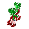
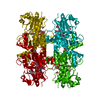
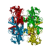
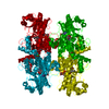
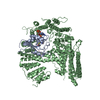
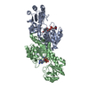
- PDB-6wkw: EM structure of CtBP2 with a minimal dehydrogenase domain of CtBP2 -
+ Open data

- Basic information
| Entry | Database: PDB / ID: 6wkw | ||||||
|---|---|---|---|---|---|---|---|
| Title | EM structure of CtBP2 with a minimal dehydrogenase domain of CtBP2 | ||||||
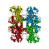
 Components Components | C-terminal-binding protein 2 | ||||||
 Keywords Keywords |  GENE REGULATION / Transcriptional corepression / Cancer / Gene repression / metabolic sensor GENE REGULATION / Transcriptional corepression / Cancer / Gene repression / metabolic sensor | ||||||
| Function / homology |  Function and homology information Function and homology informationpositive regulation of retinoic acid receptor signaling pathway / Signaling by TCF7L2 mutants / Repression of WNT target genes / Sensory processing of sound by inner hair cells of the cochlea / oxidoreductase activity, acting on the CH-OH group of donors, NAD or NADP as acceptor / white fat cell differentiation / transcription repressor complex / viral genome replication / transcription corepressor binding / transcription coregulator binding ...positive regulation of retinoic acid receptor signaling pathway / Signaling by TCF7L2 mutants / Repression of WNT target genes / Sensory processing of sound by inner hair cells of the cochlea / oxidoreductase activity, acting on the CH-OH group of donors, NAD or NADP as acceptor / white fat cell differentiation / transcription repressor complex / viral genome replication / transcription corepressor binding / transcription coregulator binding / transcription corepressor activity / NAD binding / DNA-binding transcription factor binding / transcription coactivator activity / negative regulation of cell population proliferation / negative regulation of DNA-templated transcription / synapse / protein-containing complex binding / regulation of transcription by RNA polymerase II / protein kinase binding / negative regulation of transcription by RNA polymerase II / positive regulation of transcription by RNA polymerase II / identical protein binding / nucleusSimilarity search - Function | ||||||
| Biological species |  Homo sapiens (human) Homo sapiens (human) | ||||||
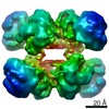
| Method | ELECTRON MICROSCOPY / single particle reconstruction / cryo EM / Resolution: 3.6 Å | ||||||
 Authors Authors | Jecrois, A.M. | ||||||
| Funding support |  United States, 1items United States, 1items
| ||||||
 Citation Citation | Journal: Structure / Year: 2021 Title: Cryo-EM structure of CtBP2 confirms tetrameric architecture. Authors: Anne M Jecrois / M Michael Dcona / Xiaoyan Deng / Dipankar Bandyopadhyay / Steven R Grossman / Celia A Schiffer / William E Royer / Abstract: C-terminal binding proteins 1 and 2 (CtBP1 and CtBP2) are transcriptional regulators that activate or repress many genes involved in cellular development, apoptosis, and metastasis. NADH-dependent ...C-terminal binding proteins 1 and 2 (CtBP1 and CtBP2) are transcriptional regulators that activate or repress many genes involved in cellular development, apoptosis, and metastasis. NADH-dependent CtBP activation has been implicated in multiple types of cancer and poor patient prognosis. Central to understanding activation of CtBP in oncogenesis is uncovering how NADH triggers protein assembly, what level of assembly occurs, and if oncogenic activity depends upon such assembly. Here, we present the cryoelectron microscopic structures of two different constructs of CtBP2 corroborating that the native state of CtBP2 in the presence of NADH is tetrameric. The physiological relevance of the observed tetramer was demonstrated in cell culture, showing that CtBP tetramer-destabilizing mutants are defective for cell migration, transcriptional repression of E-cadherin, and activation of TIAM1. Together with our cryoelectron microscopy studies, these results highlight the tetramer as the functional oligomeric form of CtBP2. | ||||||
| History |
|
- Structure visualization
| Movie |
Movie viewer |
|---|---|
| Structure viewer | Molecule: MolmilJmol/JSmol |
- Downloads & links
-Download
| PDBx/mmCIF format | 6wkw.cif.gz | 231.3 KB | Display | PDBx/mmCIF format |
|---|---|---|---|---|
| PDB format | pdb6wkw.ent.gz | 184.4 KB | Display | PDB format |
| PDBx/mmJSON format | 6wkw.json.gz | Tree view | PDBx/mmJSON format | |
| Others |  Other downloads Other downloads |
-Validation report
| Arichive directory | https://data.pdbj.org/pub/pdb/validation_reports/wk/6wkwftp://data.pdbj.org/pub/pdb/validation_reports/wk/6wkw | HTTPS FTP |
|---|
-Related structure data
| Related structure data | 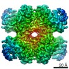 21811MC M: map data used to model this data C: citing same article ( |
|---|---|
| Similar structure data |

-Links
 PDBj
PDBj

- Assembly
| Deposited unit | 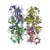
|
|---|---|
| 1 |
|
-Components
| #1: Protein | Mass: 36620.715 Da / Num. of mol.: 4 Source method: isolated from a genetically manipulated source Source: (gene. exp.) Homo sapiens (human) / Gene: CTBP2 / Production host:  Escherichia coli (E. coli) / References: UniProt: P56545 Escherichia coli (E. coli) / References: UniProt: P56545#2: Chemical | ChemComp-NAD / Nicotinamide adenine dinucleotide  Mass: 663.425 Da / Num. of mol.: 4 / Source method: obtained synthetically / Formula: C21H27N7O14P2 / Feature type: SUBJECT OF INVESTIGATION / Comment: NAD*YM Mass: 663.425 Da / Num. of mol.: 4 / Source method: obtained synthetically / Formula: C21H27N7O14P2 / Feature type: SUBJECT OF INVESTIGATION / Comment: NAD*YMHas ligand of interest | Y | |
|---|
-Experimental details
-Experiment
| Experiment | Method: ELECTRON MICROSCOPY |
|---|---|
| EM experiment | Aggregation state: PARTICLE / 3D reconstruction method: single particle reconstruction |
- Sample preparation
| Component | Name: Complex of CtBP2 with four NADH molecules / Type: COMPLEX / Entity ID: #1 / Source: RECOMBINANT |
|---|---|
| Molecular weight | Value: 0.19 MDa / Experimental value: YES |
| Source (natural) | Organism: Homo sapiens (human) |
| Source (recombinant) | Organism: Escherichia coli (E. coli) / Strain: BL21-DE3 |
| Buffer solution | pH: 7.5 |
| Specimen | Conc.: 0.25 mg/ml / Embedding applied: NO / Shadowing applied: NO / Staining applied: NO / Vitrification applied: YES |
| Specimen support | Details: Grid was washed in Ethyl acetate prior to glow-discharge. Grid material: COPPER / Grid mesh size: 400 divisions/in. / Grid type: C-flat-1.2/1.3 |
| Vitrification | Instrument: FEI VITROBOT MARK IV / Cryogen name: ETHANE / Humidity: 95 % / Chamber temperature: 278.15 K / Details: Blotting time: 4s Blotting force: 8 Wait time: 0 |
- Electron microscopy imaging
| Experimental equipment |  Model: Talos Arctica / Image courtesy: FEI Company |
|---|---|
| Microscopy | Model: FEI TALOS ARCTICA |
| Electron gun | Electron source: FIELD EMISSION GUN / Accelerating voltage: 200 kV / Illumination mode: FLOOD BEAM |
| Electron lens | Mode: BRIGHT FIELDBright-field microscopy / Nominal magnification: 50000 X |
| Specimen holder | Cryogen: NITROGEN / Specimen holder model: FEI TITAN KRIOS AUTOGRID HOLDER |
| Image recording | Average exposure time: 1.7 sec. / Electron dose: 37 e/Å2 / Film or detector model: GATAN K3 (6k x 4k) / Num. of grids imaged: 1 / Num. of real images: 3405 |
- Processing
| Software | Name: PHENIX / Version: 1.15.2_3472: / Classification: refinement | ||||||||||||||||||||||||||||||||||||||||||||
|---|---|---|---|---|---|---|---|---|---|---|---|---|---|---|---|---|---|---|---|---|---|---|---|---|---|---|---|---|---|---|---|---|---|---|---|---|---|---|---|---|---|---|---|---|---|
| EM software |
| ||||||||||||||||||||||||||||||||||||||||||||
| Image processing | Details: Images were beam-motion corrected and aligned. | ||||||||||||||||||||||||||||||||||||||||||||
| CTF correction | Type: PHASE FLIPPING AND AMPLITUDE CORRECTION | ||||||||||||||||||||||||||||||||||||||||||||
| Particle selection | Num. of particles selected: 485473 / Details: Particles were automatically picked in cisTEM. | ||||||||||||||||||||||||||||||||||||||||||||
| Symmetry | Point symmetry: D2 (2x2 fold dihedral) | ||||||||||||||||||||||||||||||||||||||||||||
| 3D reconstruction | Resolution: 3.6 Å / Resolution method: FSC 0.143 CUT-OFF / Num. of particles: 112919 / Algorithm: FOURIER SPACE / Num. of class averages: 1 / Symmetry type: POINT | ||||||||||||||||||||||||||||||||||||||||||||
| Atomic model building | Protocol: FLEXIBLE FIT | ||||||||||||||||||||||||||||||||||||||||||||
| Atomic model building | PDB-ID: 4U6Q Accession code: 4U6Q / Source name: PDB / Type: experimental model | ||||||||||||||||||||||||||||||||||||||||||||
| Refine LS restraints |
|