 Movie
Movie Controller


+ Open data

- Basic information
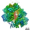
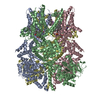
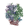
| Entry | Database: PDB / ID: 6o6b | ||||||||||||
|---|---|---|---|---|---|---|---|---|---|---|---|---|---|
| Title | Rotavirus A-VP3 (RVA-VP3) | ||||||||||||
 Components Components | Protein VP3 | ||||||||||||
 Keywords Keywords |  STRUCTURAL PROTEIN / Rotavirus / Capping enzyme / Methyl transferase / RTPase / PDE STRUCTURAL PROTEIN / Rotavirus / Capping enzyme / Methyl transferase / RTPase / PDE | ||||||||||||
| Function / homology |  Function and homology information: / Hydrolases; Acting on ester bonds; Phosphoric-diester hydrolases / mRNA guanylyltransferase activity / mRNA guanylyltransferase / mRNA (guanine-N7)-methyltransferase / viral nucleocapsid / mRNA 5'-cap (guanine-N7-)-methyltransferase activity / hydrolase activity / GTP binding / RNA binding Function and homology information: / Hydrolases; Acting on ester bonds; Phosphoric-diester hydrolases / mRNA guanylyltransferase activity / mRNA guanylyltransferase / mRNA (guanine-N7)-methyltransferase / viral nucleocapsid / mRNA 5'-cap (guanine-N7-)-methyltransferase activity / hydrolase activity / GTP binding / RNA bindingSimilarity search - Function | ||||||||||||
| Biological species |  Rotavirus A Rotavirus A | ||||||||||||
| Method | ELECTRON MICROSCOPY / single particle reconstruction / cryo EM / Resolution: 2.7 Å | ||||||||||||
 Authors Authors | Kumar, D. / Yu, X. / Prasad, V. / Wang, Z. | ||||||||||||
| Funding support |  United States, 3items United States, 3items
| ||||||||||||
 Citation Citation | Journal: To Be Published Title: A sub-atomic resolution cryo-EM of full-length Rotavirus A-VP3 (RVA-VP3) Authors: Kumar, D. / Yu, X. / Prasad, V. / Wang, Z. | ||||||||||||
| History |
|
- Structure visualization
| Movie |
Movie viewer |
|---|---|
| Structure viewer | Molecule: MolmilJmol/JSmol |
- Downloads & links
-Download
| PDBx/mmCIF format | 6o6b.cif.gz | 592.9 KB | Display | PDBx/mmCIF format |
|---|---|---|---|---|
| PDB format | pdb6o6b.ent.gz | 466 KB | Display | PDB format |
| PDBx/mmJSON format | 6o6b.json.gz | Tree view | PDBx/mmJSON format | |
| Others |  Other downloads Other downloads |
-Validation report
| Arichive directory | https://data.pdbj.org/pub/pdb/validation_reports/o6/6o6bftp://data.pdbj.org/pub/pdb/validation_reports/o6/6o6b | HTTPS FTP |
|---|
-Related structure data
| Related structure data | 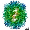 0632MC M: map data used to model this data C: citing same article ( |
|---|---|
| Similar structure data |


-Links
 PDBj
PDBj


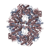
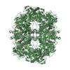
- Assembly
| Deposited unit | 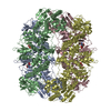
|
|---|---|
| 1 |
|
-Components
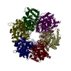
| #1: Protein | Mass: 97703.820 Da / Num. of mol.: 4 Source method: isolated from a genetically manipulated source Source: (gene. exp.) Rotavirus A / Gene: VP3 / Production host:   Spodoptera frugiperda (fall armyworm) Spodoptera frugiperda (fall armyworm)References: UniProt: Q1WK45, Hydrolases; Acting on ester bonds; Phosphoric-diester hydrolases, mRNA guanylyltransferase, mRNA (guanine-N7)-methyltransferase#2: Chemical | ChemComp-5GP / Guanosine monophosphate  Mass: 363.221 Da / Num. of mol.: 4 / Source method: obtained synthetically / Formula: C10H14N5O8P Mass: 363.221 Da / Num. of mol.: 4 / Source method: obtained synthetically / Formula: C10H14N5O8P |
|---|
-Experimental details
-Experiment
| Experiment | Method: ELECTRON MICROSCOPY |
|---|---|
| EM experiment | Aggregation state: PARTICLE / 3D reconstruction method: single particle reconstruction |
- Sample preparation
| Component | Name: VP3 / Type: COMPLEX / Entity ID: #1 / Source: RECOMBINANT |
|---|---|
| Source (natural) | Organism: Rotavirus A |
| Source (recombinant) | Organism: Spodoptera frugiperda (fall armyworm) |
| Details of virus | Empty: NO / Enveloped: NO / Isolate: SPECIES / Type: PRION |
| Buffer solution | pH: 7 |
| Specimen | Conc.: 0.5 mg/ml / Embedding applied: NO / Shadowing applied: NO / Staining applied: NO / Vitrification applied: YES |
| Specimen support | Grid material: COPPER / Grid mesh size: 400 divisions/in. / Grid type: Quantifoil R1.2/1.3 |
| Vitrification | Instrument: FEI VITROBOT MARK IV / Cryogen name: ETHANE / Humidity: 95 % / Chamber temperature: 295 K |
- Electron microscopy imaging
| Microscopy | Model: JEOL 3200FSC |
|---|---|
| Electron gun | Electron source: FIELD EMISSION GUN / Accelerating voltage: 300 kV / Illumination mode: FLOOD BEAM |
| Electron lens | Mode: BRIGHT FIELDBright-field microscopy / Nominal magnification: 40000 X / Nominal defocus max: 2000 nm / Nominal defocus min: 1000 nm / Cs: 4.7 mm / C2 aperture diameter: 100 µm |
| Image recording | Average exposure time: 10 sec. / Electron dose: 50 e/Å2 / Detector mode: SUPER-RESOLUTION / Film or detector model: GATAN K2 SUMMIT (4k x 4k) / Num. of grids imaged: 1 |
| Image scans | Width: 7420 / Height: 7676 / Movie frames/image: 50 |
- Processing
| EM software |
| ||||||||||||||||||||||||||||||||||||||||
|---|---|---|---|---|---|---|---|---|---|---|---|---|---|---|---|---|---|---|---|---|---|---|---|---|---|---|---|---|---|---|---|---|---|---|---|---|---|---|---|---|---|
| CTF correction | Type: PHASE FLIPPING ONLY | ||||||||||||||||||||||||||||||||||||||||
| Particle selection | Num. of particles selected: 133712 | ||||||||||||||||||||||||||||||||||||||||
| Symmetry | Point symmetry: D2 (2x2 fold dihedral) | ||||||||||||||||||||||||||||||||||||||||
| 3D reconstruction | Resolution: 2.7 Å / Resolution method: FSC 0.143 CUT-OFF / Num. of particles: 70892 / Symmetry type: POINT |
