[English] 日本語
Yorodumi
- PDB-6o3v: Crystal structure for RVA-VP3 -

+
Open data


ID or keywords:

Loading...

-
Basic information

Entry
Database: PDB / ID: 6o3v
TitleCrystal structure for RVA-VP3
ComponentsProtein VP3
KeywordsSTRUCTURAL PROTEIN / Rotavirus / VP3 / Capping enzyme / Guanylyl transferase / Methyl transferase / RTPase / PDE
Function / homology
Function and homology information


Hydrolases; Acting on ester bonds; Phosphoric-diester hydrolases / : / mRNA guanylyltransferase activity / mRNA guanylyltransferase / mRNA (guanine-N7)-methyltransferase / viral nucleocapsid / mRNA 5'-cap (guanine-N7-)-methyltransferase activity / hydrolase activity / GTP binding / RNA binding
Similarity search - Function
Protein VP3, rotavirus / Rotavirus VP3 protein / Viral protein 3 containing mRNA (guanine-N(7)-)-methyltransferase family profile.
Similarity search - Domain/homology
GUANOSINE-5'-MONOPHOSPHATE / Protein VP3
Similarity search - Component
Biological speciesRotavirus A
MethodX-RAY DIFFRACTION / SYNCHROTRON / MOLECULAR REPLACEMENT / Resolution: 3.5 Å
AuthorsKumar, D. / Yu, X. / Wang, Z. / Hu, L. / Prasad, V.
Funding support United States, 2items
OrganizationGrant numberCountry
National Institutes of Health/National Institute Of Allergy and Infectious Diseases (NIH/NIAID)AI36040 United States
Welch FoundationQ1279 United States
Citation
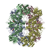
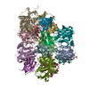
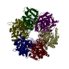
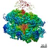
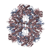
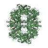
Journal: Sci Adv / Year: 2020
Title: 2.7 Å cryo-EM structure of rotavirus core protein VP3, a unique capping machine with a helicase activity.
Authors: Dilip Kumar / Xinzhe Yu / Sue E Crawford / Rodolfo Moreno / Joanita Jakana / Banumathi Sankaran / Ramakrishnan Anish / Soni Kaundal / Liya Hu / Mary K Estes / Zhao Wang / B V Venkataram Prasad /
Abstract: In many viruses, including rotavirus (RV), the major pathogen of infantile gastroenteritis, capping of viral messenger RNAs is a pivotal step for efficient translation of the viral genome. In RV, VP3 ...In many viruses, including rotavirus (RV), the major pathogen of infantile gastroenteritis, capping of viral messenger RNAs is a pivotal step for efficient translation of the viral genome. In RV, VP3 caps the nascent transcripts synthesized from the genomic dsRNA segments by the RV polymerase VP1 within the particle core. Here, from cryo-electron microscopy, x-ray crystallography, and biochemical analyses, we show that VP3 forms a stable tetrameric assembly with each subunit having a modular domain organization, which uniquely integrates five distinct enzymatic steps required for capping the transcripts. In addition to the previously known guanylyl- and methyltransferase activities, we show that VP3 exhibits hitherto unsuspected RNA triphosphatase activity necessary for initiating transcript capping and RNA helicase activity likely required for separating the RNA duplex formed transiently during endogenous transcription. From our studies, we propose a new mechanism for how VP3 inside the virion core caps the nascent transcripts exiting from the polymerase.
History
DepositionFeb 27, 2019Deposition site: RCSB / Processing site: RCSB
Revision 1.0Mar 4, 2020Provider: repository / Type: Initial release
Revision 1.1Sep 16, 2020Group: Database references / Category: citation / citation_author
Item: _citation.country / _citation.journal_abbrev ..._citation.country / _citation.journal_abbrev / _citation.journal_id_CSD / _citation.journal_id_ISSN / _citation.journal_volume / _citation.page_first / _citation.page_last / _citation.pdbx_database_id_DOI / _citation.pdbx_database_id_PubMed / _citation.title / _citation.year
Revision 1.2Mar 13, 2024Group: Data collection / Database references / Category: chem_comp_atom / chem_comp_bond / database_2
Item: _database_2.pdbx_DOI / _database_2.pdbx_database_accession

-
Structure visualization

Structure viewerMolecule:
MolmilJmol/JSmol

Downloads & links

-
Assembly

Deposited unit
A: Protein VP3
B: Protein VP3
C: Protein VP3
hetero molecules


Theoretical massNumber of molelcules
Total (without water)294,67612
Polymers293,1113
Non-polymers1,5649
Water48627
1
A: Protein VP3
C: Protein VP3
hetero molecules

A: Protein VP3
C: Protein VP3
hetero molecules


  • defined by author&software
  • Evidence: gel filtration, Gel filtration profile indicates a tetrameric assembly in solution, equilibrium centrifugation, Analytical Ultracentrifugation confirms tetrameric state is preferred ...Evidence: gel filtration, Gel filtration profile indicates a tetrameric assembly in solution, equilibrium centrifugation, Analytical Ultracentrifugation confirms tetrameric state is preferred oligomeric assembly in solution
  • 393 kDa, 4 polymers
  • Search similar-shape structures of this assembly by Omokage search (details)
Theoretical massNumber of molelcules
Total (without water)392,90116
Polymers390,8154
Non-polymers2,08512
Water724
TypeNameSymmetry operationNumber
identity operation1_555x,y,z1
crystal symmetry operation11_555-x+y,y,-z1
Buried area23160 Å2
ΔGint-146 kcal/mol
Surface area126700 Å2
MethodPISA
2
B: Protein VP3
hetero molecules

B: Protein VP3
hetero molecules

B: Protein VP3
hetero molecules

B: Protein VP3
hetero molecules


Theoretical massNumber of molelcules
Total (without water)392,90116
Polymers390,8154
Non-polymers2,08512
Water724
TypeNameSymmetry operationNumber
identity operation1_555x,y,z1
crystal symmetry operation4_545-x,-y-1,z1
crystal symmetry operation9_555-x,-x+y,-z+1/31
crystal symmetry operation12_545x,x-y-1,-z+1/31
Buried area23840 Å2
ΔGint-163 kcal/mol
Surface area130580 Å2
MethodPISA
Unit cell
Length a, b, c (Å)236.706, 236.706, 349.300
Angle α, β, γ (deg.)90.00, 90.00, 120.00
Int Tables number180
Space group name H-MP6222

-
Components

#1: Protein Protein VP3


Mass: 97703.820 Da / Num. of mol.: 3
Source method: isolated from a genetically manipulated source
Source: (gene. exp.) Rotavirus A / Gene: VP3 / Production host: Spodoptera frugiperda (fall armyworm) / References: UniProt: Q1WK45
#2: Chemical ChemComp-5GP / GUANOSINE-5'-MONOPHOSPHATE / Guanosine monophosphate


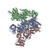
Mass: 363.221 Da / Num. of mol.: 3 / Source method: obtained synthetically / Formula: C10H14N5O8P
#3: Chemical ChemComp-EDO / 1,2-ETHANEDIOL / ETHYLENE GLYCOL / Ethylene glycol


Mass: 62.068 Da / Num. of mol.: 3 / Source method: obtained synthetically / Formula: C2H6O2
#4: Chemical ChemComp-SO4 / SULFATE ION / Sulfate


Mass: 96.063 Da / Num. of mol.: 3 / Source method: obtained synthetically / Formula: SO4
#5: Water ChemComp-HOH / water / Water


Mass: 18.015 Da / Num. of mol.: 27 / Source method: isolated from a natural source / Formula: H2O

-
Experimental details

-
Experiment

ExperimentMethod: X-RAY DIFFRACTION / Number of used crystals: 1

-
Sample preparation

CrystalDensity Matthews: 4.82 Å3/Da / Density % sol: 74.47 %
Crystal growTemperature: 293.15 K / Method: vapor diffusion, hanging drop / pH: 5.5
Details: Protein conc ~ 7 mg/ml 0.2 M Lithium Sulphate 0.1 M Sodium citrate pH 5.5 15% Ethanol

-
Data collection

DiffractionMean temperature: 80 K / Serial crystal experiment: N
Diffraction sourceSource: SYNCHROTRON / Site: ALS / Beamline: 5.0.2 / Wavelength: 0.9999 Å
DetectorType: ADSC QUANTUM 315r / Detector: CCD / Date: Aug 16, 2017
RadiationProtocol: SINGLE WAVELENGTH / Monochromatic (M) / Laue (L): M / Scattering type: x-ray
Radiation wavelengthWavelength: 0.9999 Å / Relative weight: 1
ReflectionResolution: 3.5→49.175 Å / Num. obs: 71574 / % possible obs: 97.67 % / Redundancy: 14.7 % / Rpim(I) all: 0.052 / Net I/σ(I): 4.5
Reflection shellResolution: 3.5→3.625 Å / Num. unique obs: 5807

-
Processing

Software
NameVersionClassification
PHENIX(1.14_3235: ???)refinement
HKL-2000data reduction
HKL-2000data scaling
PHASERphasing
RefinementMethod to determine structure: MOLECULAR REPLACEMENT / Resolution: 3.5→49.175 Å / SU ML: 0.4 / Cross valid method: FREE R-VALUE / σ(F): 1.33 / Phase error: 25.38
RfactorNum. reflection% reflection
Rfree0.2644 3499 4.9 %
Rwork0.229 --
obs0.2307 71431 97.74 %
Solvent computationShrinkage radii: 0.9 Å / VDW probe radii: 1.11 Å
Refinement stepCycle: LAST / Resolution: 3.5→49.175 Å
ProteinNucleic acidLigandSolventTotal
Num. atoms20604 0 99 27 20730
Refine LS restraints
Refine-IDTypeDev idealNumber
X-RAY DIFFRACTIONf_bond_d0.00521225
X-RAY DIFFRACTIONf_angle_d0.87128810
X-RAY DIFFRACTIONf_dihedral_angle_d7.06912503
X-RAY DIFFRACTIONf_chiral_restr0.0533164
X-RAY DIFFRACTIONf_plane_restr0.0053614
LS refinement shell
Resolution (Å)Rfactor RfreeNum. reflection RfreeRfactor RworkNum. reflection RworkRefine-ID% reflection obs (%)
3.5-3.54790.32271110.29341895X-RAY DIFFRACTION70
3.5479-3.59860.31961080.29752342X-RAY DIFFRACTION85
3.5986-3.65230.4351350.39332556X-RAY DIFFRACTION94
3.6523-3.70940.46221310.38232665X-RAY DIFFRACTION98
3.7094-3.77020.27891560.27872736X-RAY DIFFRACTION100
3.7702-3.83510.28441510.26632704X-RAY DIFFRACTION100
3.8351-3.90480.33191270.30132745X-RAY DIFFRACTION100
3.9048-3.97990.31481470.2962713X-RAY DIFFRACTION99
3.9799-4.06110.2731490.24522747X-RAY DIFFRACTION100
4.0611-4.14940.23431240.23482752X-RAY DIFFRACTION100
4.1494-4.24590.26221260.22152751X-RAY DIFFRACTION100
4.2459-4.3520.25281570.2152735X-RAY DIFFRACTION100
4.352-4.46960.23871540.20522744X-RAY DIFFRACTION100
4.4696-4.6010.241480.19882746X-RAY DIFFRACTION100
4.601-4.74940.24211330.18862761X-RAY DIFFRACTION100
4.7494-4.9190.23111570.19422741X-RAY DIFFRACTION100
4.919-5.11570.23871420.1962771X-RAY DIFFRACTION100
5.1157-5.34830.23331270.19862806X-RAY DIFFRACTION100
5.3483-5.62990.24861590.20732773X-RAY DIFFRACTION100
5.6299-5.98210.23581640.21662773X-RAY DIFFRACTION100
5.9821-6.4430.241280.21992818X-RAY DIFFRACTION100
6.443-7.08970.26041390.2222835X-RAY DIFFRACTION100
7.0897-8.11160.20361460.19962853X-RAY DIFFRACTION100
8.1116-10.20470.20651260.16222914X-RAY DIFFRACTION100
10.2047-49.17980.25331540.20043056X-RAY DIFFRACTION99
Refinement TLS params.

Method: refined / Refine-ID: X-RAY DIFFRACTION

IDL112)L122)L132)L222)L232)L332)S11 (Å °)S12 (Å °)S13 (Å °)S21 (Å °)S22 (Å °)S23 (Å °)S31 (Å °)S32 (Å °)S33 (Å °)T112)T122)T132)T222)T232)T332)Origin x (Å)Origin y (Å)Origin z (Å)
10.3166-0.44560.04310.6623-0.19110.4319-0.0468-0.08450.09870.24730.037-0.1859-0.09430.00930.09130.81540.2879-0.46970.2884-0.15780.470224.2893-56.578133.2891
20.45860.00540.11590.9111-0.15330.1447-0.01640.18-0.0022-0.22550.097-0.70140.25910.2119-0.05340.86190.2503-0.23250.3148-0.32320.776935.5199-65.7548-3.8384
30.5611-0.55690.19320.9181-0.37130.3995-0.0868-0.1519-0.05610.18220.20670.13060.12640.0101-0.01830.72050.23-0.36120.2046-0.030.36245.5908-62.883122.4132
40.35490.2677-0.10470.4925-0.70261.38450.0342-0.4828-0.27280.72540.03060.16180.0558-0.1369-0.21391.22270.132-0.16230.47120.25460.4778-14.6132-72.90938.4819
50.4048-0.31890.3750.7051-0.62330.70910.0122-0.0858-0.0681-0.05420.09340.1928-0.0403-0.15610.02660.76930.4325-0.3530.3149-0.10310.292930.6576-73.892148.7653
60.4549-0.6639-0.04561.0576-0.06770.64830.17130.2739-0.7736-0.5797-0.13961.07590.3444-0.43010.07631.08290.2489-0.62930.452-0.22481.230124.247-109.962537.8575
70.7519-0.3220.44040.5720.09290.42770.0650.06050.076-0.201-0.0405-0.0851-0.05320.00450.16290.94240.3835-0.19260.2918-0.06990.259351.5954-80.561641.1637
80.42350.01840.27861.5832-0.31390.33860.16750.32220.8611-0.4636-0.1244-0.6181-0.55190.15250.18121.34990.36190.30120.41670.25740.959768.8306-63.876231.7231
90.46620.33740.0961.6193-0.1440.40110.0425-0.25380.07150.67010.14290.28640.258-0.0265-0.11870.83040.307-0.14420.3742-0.12630.5069-5.2643-35.652335.5688
101.0034-0.34760.2220.9708-0.17560.658-0.0982-0.1590.23940.40260.24440.42430.0522-0.1673-0.10720.49830.1356-0.03010.21530.01550.7669-22.524-22.694121.0248
111.4389-0.9432-0.52111.8613-0.09950.34590.0223-0.34580.29130.38710.2115-0.6227-0.05610.229-0.19630.43920.1199-0.29240.3237-0.26210.69124.0946-27.68817.9456
120.2169-0.09380.09550.25990.03760.2789-0.0459-0.07180.16880.26070.1082-0.68780.06310.2997-0.15690.42440.1466-0.39820.3441-0.26071.152140.6306-19.342313.1381
Refinement TLS group
IDRefine-IDRefine TLS-IDSelection details
1X-RAY DIFFRACTION1chain 'A' and (resid 1 through 94 )
2X-RAY DIFFRACTION2chain 'A' and (resid 95 through 498 )
3X-RAY DIFFRACTION3chain 'A' and (resid 499 through 627 )
4X-RAY DIFFRACTION4chain 'A' and (resid 628 through 835 )
5X-RAY DIFFRACTION5chain 'B' and (resid 1 through 104 )
6X-RAY DIFFRACTION6chain 'B' and (resid 105 through 498 )
7X-RAY DIFFRACTION7chain 'B' and (resid 499 through 618 )
8X-RAY DIFFRACTION8chain 'B' and (resid 619 through 835 )
9X-RAY DIFFRACTION9chain 'C' and (resid 1 through 217 )
10X-RAY DIFFRACTION10chain 'C' and (resid 218 through 579 )
11X-RAY DIFFRACTION11chain 'C' and (resid 580 through 651 )
12X-RAY DIFFRACTION12chain 'C' and (resid 652 through 835 )

+
About Yorodumi

-
News

-
Feb 9, 2022. New format data for meta-information of EMDB entries

New format data for meta-information of EMDB entries

  • Version 3 of the EMDB header file is now the official format.
  • The previous official version 1.9 will be removed from the archive.

Related info.:EMDB header

External links:wwPDB to switch to version 3 of the EMDB data model

-
Aug 12, 2020. Covid-19 info

Covid-19 info

URL: https://pdbj.org/emnavi/covid19.php

New page: Covid-19 featured information page in EM Navigator.

Related info.:Covid-19 info / Mar 5, 2020. Novel coronavirus structure data

+
Mar 5, 2020. Novel coronavirus structure data

Novel coronavirus structure data

Related info.:Yorodumi Speices / Aug 12, 2020. Covid-19 info

External links:COVID-19 featured content - PDBj / Molecule of the Month (242):Coronavirus Proteases

+
Jan 31, 2019. EMDB accession codes are about to change! (news from PDBe EMDB page)

EMDB accession codes are about to change! (news from PDBe EMDB page)

  • The allocation of 4 digits for EMDB accession codes will soon come to an end. Whilst these codes will remain in use, new EMDB accession codes will include an additional digit and will expand incrementally as the available range of codes is exhausted. The current 4-digit format prefixed with “EMD-” (i.e. EMD-XXXX) will advance to a 5-digit format (i.e. EMD-XXXXX), and so on. It is currently estimated that the 4-digit codes will be depleted around Spring 2019, at which point the 5-digit format will come into force.
  • The EM Navigator/Yorodumi systems omit the EMD- prefix.

Related info.:Q: What is EMD? / ID/Accession-code notation in Yorodumi/EM Navigator

External links:EMDB Accession Codes are Changing Soon! / Contact to PDBj

+
Jul 12, 2017. Major update of PDB

Major update of PDB

  • wwPDB released updated PDB data conforming to the new PDBx/mmCIF dictionary.
  • This is a major update changing the version number from 4 to 5, and with Remediation, in which all the entries are updated.
  • In this update, many items about electron microscopy experimental information are reorganized (e.g. em_software).
  • Now, EM Navigator and Yorodumi are based on the updated data.

External links:wwPDB Remediation / Enriched Model Files Conforming to OneDep Data Standards Now Available in the PDB FTP Archive

-
Yorodumi

Thousand views of thousand structures

  • Yorodumi is a browser for structure data from EMDB, PDB, SASBDB, etc.
  • This page is also the successor to EM Navigator detail page, and also detail information page/front-end page for Omokage search.
  • The word "yorodu" (or yorozu) is an old Japanese word meaning "ten thousand". "mi" (miru) is to see.

Related info.:EMDB / PDB / SASBDB / Comparison of 3 databanks / Yorodumi Search / Aug 31, 2016. New EM Navigator & Yorodumi / Yorodumi Papers / Jmol/JSmol / Function and homology information / Changes in new EM Navigator and Yorodumi

Read more