 Movie
Movie Controller


+ Open data

- Basic information
| Entry | Database: PDB / ID: 3k2r | ||||||
|---|---|---|---|---|---|---|---|
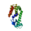
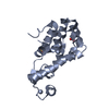
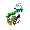
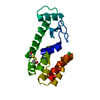
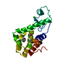
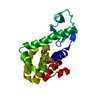
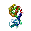
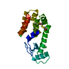
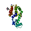
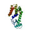
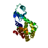
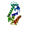
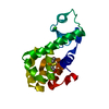
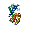
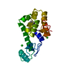
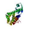
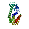
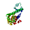
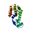
| Title | Crystal Structure of Spin Labeled T4 Lysozyme Mutant K65V1/R76V1 | ||||||
 Components Components | Lysozyme | ||||||
 Keywords Keywords | HYDROLASE / NITROXIDE SPIN LABEL / EPR / MODIFIED CYSTEINE / Antimicrobial / Bacteriolytic enzyme / Glycosidase | ||||||
| Function / homology |  Function and homology information Function and homology informationviral release from host cell by cytolysis / peptidoglycan catabolic process / cell wall macromolecule catabolic process / lysozyme / lysozyme activity / host cell cytoplasm / defense response to bacteriumSimilarity search - Function | ||||||
| Biological species |  Enterobacteria phage T4 (virus) Enterobacteria phage T4 (virus) | ||||||
| Method | X-RAY DIFFRACTION / MOLECULAR REPLACEMENT / molecular replacement / Resolution: 1.5 Å | ||||||
 Authors Authors | Toledo Warshaviak, D. / Cascio, D. / Khramtsov, V.V. / Hubbell, W.L. | ||||||
 Citation Citation | Journal: To be Published Title: Crystal Structure of Spin Labeled T4 Lysozyme Mutant K65V1/R76V1 Authors: Toledo Warshaviak, D. / Cascio, D. / Khramtsov, V.V. / Hubbell, W.L. | ||||||
| History |
|
- Structure visualization
| Structure viewer | Molecule: MolmilJmol/JSmol |
|---|
- Downloads & links
-Download
| PDBx/mmCIF format | 3k2r.cif.gz | 51.3 KB | Display | PDBx/mmCIF format |
|---|---|---|---|---|
| PDB format | pdb3k2r.ent.gz | 34.9 KB | Display | PDB format |
| PDBx/mmJSON format | 3k2r.json.gz | Tree view | PDBx/mmJSON format | |
| Others |  Other downloads Other downloads |
-Validation report
| Arichive directory | https://data.pdbj.org/pub/pdb/validation_reports/k2/3k2rftp://data.pdbj.org/pub/pdb/validation_reports/k2/3k2r | HTTPS FTP |
|---|
-Related structure data
| Related structure data | 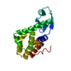 3lzmS S: Starting model for refinement |
|---|---|
| Similar structure data |

-Links
 PDBj
PDBj


- Assembly
| Deposited unit | 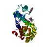
| ||||||||
|---|---|---|---|---|---|---|---|---|---|
| 1 |
| ||||||||
| Unit cell |
|
-Components
-Protein , 1 types, 1 molecules A
| #1: Protein | / Lysis protein / Muramidase / Endolysin Mass: 18548.273 Da / Num. of mol.: 1 / Mutation: C54T,K65C,R76C,C97A Source method: isolated from a genetically manipulated source Source: (gene. exp.) Enterobacteria phage T4 (virus) / Gene: E, Lysozyme / Plasmid: pHSe5 / Production host:  Escherichia coli (E. coli) / Strain (production host): BL21 / References: UniProt: P00720, lysozyme Escherichia coli (E. coli) / Strain (production host): BL21 / References: UniProt: P00720, lysozyme |
|---|
-Non-polymers , 5 types, 156 molecules 


| #2: Chemical |  Mass: 251.346 Da / Num. of mol.: 2 / Source method: obtained synthetically / Formula: C8H15N2O3S2 Mass: 251.346 Da / Num. of mol.: 2 / Source method: obtained synthetically / Formula: C8H15N2O3S2#3: Chemical | 1,6-Hexanediol Mass: 118.174 Da / Num. of mol.: 2 / Source method: obtained synthetically / Formula: C6H14O2 Mass: 118.174 Da / Num. of mol.: 2 / Source method: obtained synthetically / Formula: C6H14O2#4: Chemical | Chloride Mass: 35.453 Da / Num. of mol.: 2 / Source method: obtained synthetically / Formula: Cl Mass: 35.453 Da / Num. of mol.: 2 / Source method: obtained synthetically / Formula: Cl#5: Chemical | ChemComp-K / |  Mass: 39.098 Da / Num. of mol.: 1 / Source method: obtained synthetically / Formula: K Mass: 39.098 Da / Num. of mol.: 1 / Source method: obtained synthetically / Formula: K#6: Water | ChemComp-HOH / | WaterMass: 18.015 Da / Num. of mol.: 149 / Source method: isolated from a natural source / Formula: H2O |
|---|
-Experimental details
-Experiment
| Experiment | Method: X-RAY DIFFRACTION / Number of used crystals: 1 |
|---|
- Sample preparation
| Crystal | Density Matthews: 2.63 Å3/Da / Density % sol: 53.21 % |
|---|---|
| Crystal grow | Temperature: 293 K / Method: vapor diffusion, hanging drop / pH: 7 Details: 2.2M dibasic potassium phosphate and monobasic sodium phosphate, 0.15M sodium chloride, 100mM 1,6 hexanediol, pH 7.0, vapor diffusion, hanging drop, temperature 293K |
-Data collection
| Diffraction | Mean temperature: 100 K |
|---|---|
| Diffraction source | Source: ROTATING ANODE / Type: RIGAKU FR-D / Wavelength: 1.5418 Å |
| Detector | Type: RIGAKU RAXIS IV++ / Detector: IMAGE PLATE / Date: Feb 4, 2008 / Details: CONFOCAL MIRRORS (BLUE OPTICS) |
| Radiation | Monochromator: CONFOCAL MIRRORS (BLUE OPTICS) / Protocol: SINGLE WAVELENGTH / Monochromatic (M) / Laue (L): M / Scattering type: x-ray |
| Radiation wavelength | Wavelength: 1.5418 Å / Relative weight: 1 |
| Reflection | Resolution: 1.5→80 Å / Num. obs: 30957 / % possible obs: 96.8 % / Redundancy: 5.7 % / Biso Wilson estimate: 20.8 Å2 / Rmerge(I) obs: 0.058 / Χ2: 1.074 / Net I/σ(I): 20.4 |
| Reflection shell | Resolution: 1.5→1.55 Å / Redundancy: 2.4 % / Rmerge(I) obs: 0.368 / Mean I/σ(I) obs: 2.16 / Num. unique all: 2395 / Χ2: 0.897 / % possible all: 76.2 |
-Phasing
| Phasing | Method: molecular replacement |
|---|
- Processing
| Software |
| ||||||||||||||||||||||||||||||||||||||||||||||||||||||||||||||||||||||||||||||||||||
|---|---|---|---|---|---|---|---|---|---|---|---|---|---|---|---|---|---|---|---|---|---|---|---|---|---|---|---|---|---|---|---|---|---|---|---|---|---|---|---|---|---|---|---|---|---|---|---|---|---|---|---|---|---|---|---|---|---|---|---|---|---|---|---|---|---|---|---|---|---|---|---|---|---|---|---|---|---|---|---|---|---|---|---|---|---|
| Refinement | Method to determine structure: MOLECULAR REPLACEMENT Starting model: PDB ENTRY 3LZM Resolution: 1.5→25.248 Å / Occupancy max: 1 / Occupancy min: 0 / FOM work R set: 0.885 / SU ML: 0.16 / Cross valid method: THROUGHOUT / σ(F): 1.36 / Stereochemistry target values: ML
| ||||||||||||||||||||||||||||||||||||||||||||||||||||||||||||||||||||||||||||||||||||
| Solvent computation | Shrinkage radii: 0.9 Å / VDW probe radii: 1.11 Å / Solvent model: FLAT BULK SOLVENT MODEL / Bsol: 42.933 Å2 / ksol: 0.392 e/Å3 | ||||||||||||||||||||||||||||||||||||||||||||||||||||||||||||||||||||||||||||||||||||
| Displacement parameters | Biso max: 62.13 Å2 / Biso mean: 19.139 Å2 / Biso min: 11.71 Å2
| ||||||||||||||||||||||||||||||||||||||||||||||||||||||||||||||||||||||||||||||||||||
| Refinement step | Cycle: LAST / Resolution: 1.5→25.248 Å
| ||||||||||||||||||||||||||||||||||||||||||||||||||||||||||||||||||||||||||||||||||||
| Refine LS restraints |
| ||||||||||||||||||||||||||||||||||||||||||||||||||||||||||||||||||||||||||||||||||||
| LS refinement shell | Refine-ID: X-RAY DIFFRACTION / Total num. of bins used: 11
|
