 Movie
Movie Controller

[English] 日本語
 Yorodumi
- PDB-1aaw: THE STRUCTURAL BASIS FOR THE ALTERED SUBSTRATE SPECIFICITY OF THE... -
+ Open data

- Basic information
| Entry | Database: PDB / ID: 1aaw | ||||||
|---|---|---|---|---|---|---|---|
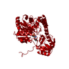
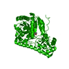
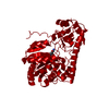
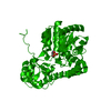
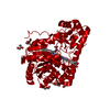
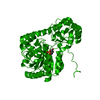
| Title | THE STRUCTURAL BASIS FOR THE ALTERED SUBSTRATE SPECIFICITY OF THE R292D ACTIVE SITE MUTANT OF ASPARTATE AMINOTRANSFERASE FROM E. COLI | ||||||
 Components Components | ASPARTATE AMINOTRANSFERASE | ||||||
 Keywords Keywords | AMINOTRANSFERASE | ||||||
| Function / homology |  Function and homology information Function and homology informationL-phenylalanine biosynthetic process from chorismate via phenylpyruvate / L-tyrosine-2-oxoglutarate transaminase activity / L-phenylalanine biosynthetic process / aspartate transaminase / L-aspartate:2-oxoglutarate aminotransferase activity / pyridoxal phosphate binding / protein homodimerization activity / identical protein binding / cytoplasm / cytosol Similarity search - Function | ||||||
| Biological species |  | ||||||
| Method |  X-RAY DIFFRACTION / Resolution: 2.4 Å X-RAY DIFFRACTION / Resolution: 2.4 Å | ||||||
 Authors Authors | Almo, S.C. / Smith, D.L. / Danishefsky, A.T. / Ringe, D. | ||||||
 Citation Citation | Journal: Protein Eng. / Year: 1994 Title: The structural basis for the altered substrate specificity of the R292D active site mutant of aspartate aminotransferase from E. coli. Authors: Almo, S.C. / Smith, D.L. / Danishefsky, A.T. / Ringe, D. #1: Journal: Biochemistry / Year: 1991Title: Activity and Structure of the Active Site Mutants R386Y and R386F of Escherichia Coli Aspartate Aminotransferase Authors: Danishefsky, A.T. / Onnufer, J.J. / Petsko, G.A. / Ringe, D. #2: Journal: Biochemistry / Year: 1989Title: 2.8 Angstroms Resolution Crystal Structure of an Active Site Mutant of Aspartate Aminotransferase from Escherichia Coli Authors: Smith, D.L. / Almo, S.C. / Toney, M.D. / Ringe, D. | ||||||
| History |
|
- Structure visualization
| Structure viewer | Molecule: MolmilJmol/JSmol |
|---|
- Downloads & links
-Download
| PDBx/mmCIF format | 1aaw.cif.gz | 82.7 KB | Display | PDBx/mmCIF format |
|---|---|---|---|---|
| PDB format | pdb1aaw.ent.gz | 62.7 KB | Display | PDB format |
| PDBx/mmJSON format | 1aaw.json.gz | Tree view | PDBx/mmJSON format | |
| Others |  Other downloads Other downloads |
-Validation report
| Summary document | 1aaw_validation.pdf.gz | 389.8 KB | Display | wwPDB validaton report |
|---|---|---|---|---|
| Full document | 1aaw_full_validation.pdf.gz | 410.1 KB | Display | |
| Data in XML | 1aaw_validation.xml.gz | 12.1 KB | Display | |
| Data in CIF | 1aaw_validation.cif.gz | 17.2 KB | Display | |
| Arichive directory | https://data.pdbj.org/pub/pdb/validation_reports/aa/1aawftp://data.pdbj.org/pub/pdb/validation_reports/aa/1aaw | HTTPS FTP |
-Related structure data


-Links
 PDBj
PDBj- Assembly
| Deposited unit | 
| ||||||||
|---|---|---|---|---|---|---|---|---|---|
| 1 | 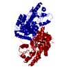
| ||||||||
| Unit cell |
| ||||||||
| Atom site foot note | 1: ALA 20 - PRO 21 OMEGA ANGLE = 258.164 PEPTIDE BOND DEVIATES SIGNIFICANTLY FROM TRANS CONFORMATION 2: CIS PROLINE - PRO 140 / 3: CIS PROLINE - PRO 196 4: ASP 200 - PRO 201 OMEGA ANGLE = 115.757 PEPTIDE BOND DEVIATES SIGNIFICANTLY FROM TRANS CONFORMATION 5: RESIDUE PLP 409 IS BOUND TO LYS 258 FORMING A PROTONATED SCHIFF BASE LINKAGE BETWEEN NZ LYS 258 AND C4A PLP 409. | ||||||||
| Details | THE MOLECULE IS A DIMER. TO GENERATE THE OTHER CHAIN ONE MUST APPLY THE CRYSTALLOGRAPHIC SYMMETRY OPERATION (X, 86.9-Y, 80.0-Z) TO THE COORDINATES IN THIS ENTRY. |
-Components
| #1: Protein | Mass: 43619.215 Da / Num. of mol.: 1 Source method: isolated from a genetically manipulated source Source: (gene. exp.) |
|---|---|
| #2: Chemical | ChemComp-PLP /   Mass: 247.142 Da / Num. of mol.: 1 / Source method: obtained synthetically / Formula: C8H10NO6P Mass: 247.142 Da / Num. of mol.: 1 / Source method: obtained synthetically / Formula: C8H10NO6P |
| #3: Water | ChemComp-HOH /  Mass: 18.015 Da / Num. of mol.: 3 / Source method: isolated from a natural source / Formula: H2O Mass: 18.015 Da / Num. of mol.: 3 / Source method: isolated from a natural source / Formula: H2O |
-Experimental details
-Experiment
| Experiment | Method: X-RAY DIFFRACTION |
|---|
- Sample preparation
| Crystal | Density Matthews: 3.12 Å3/Da / Density % sol: 60.61 % | |||||||||||||||||||||||||
|---|---|---|---|---|---|---|---|---|---|---|---|---|---|---|---|---|---|---|---|---|---|---|---|---|---|---|
| Crystal grow | *PLUS pH: 7.5 / Method: vapor diffusion / Details: referred to J.Mol.Biol. 191.301-302 1986 | |||||||||||||||||||||||||
| Components of the solutions | *PLUS
|
-Data collection
| Radiation | Scattering type: x-ray |
|---|---|
| Radiation wavelength | Relative weight: 1 |
| Reflection | *PLUS Highest resolution: 2.4 Å / Num. obs: 15069 / % possible obs: 72 % |
- Processing
| Software |
| |||||||||||||||||||||||||||||||||||||||||||||||||||||||||||||||
|---|---|---|---|---|---|---|---|---|---|---|---|---|---|---|---|---|---|---|---|---|---|---|---|---|---|---|---|---|---|---|---|---|---|---|---|---|---|---|---|---|---|---|---|---|---|---|---|---|---|---|---|---|---|---|---|---|---|---|---|---|---|---|---|---|
| Refinement | Rfactor obs: 0.208 / Highest resolution: 2.4 Å | |||||||||||||||||||||||||||||||||||||||||||||||||||||||||||||||
| Refinement step | Cycle: LAST / Highest resolution: 2.4 Å
| |||||||||||||||||||||||||||||||||||||||||||||||||||||||||||||||
| Refine LS restraints |
| |||||||||||||||||||||||||||||||||||||||||||||||||||||||||||||||
| Refinement | *PLUS Highest resolution: 2.4 Å / Lowest resolution: 5 Å / Num. reflection obs: 15069 / σ(I): 2 / Rfactor obs: 0.208 | |||||||||||||||||||||||||||||||||||||||||||||||||||||||||||||||
| Solvent computation | *PLUS | |||||||||||||||||||||||||||||||||||||||||||||||||||||||||||||||
| Displacement parameters | *PLUS Biso mean: 15 Å2 | |||||||||||||||||||||||||||||||||||||||||||||||||||||||||||||||
| Refine LS restraints | *PLUS
|
