 Movie
Movie Controller

[English] 日本語
 Yorodumi
- PDB-1cr2: CRYSTAL STRUCTURE OF THE HELICASE DOMAIN OF THE GENE 4 PROTEIN OF... -
+ Open data

- Basic information
| Entry | Database: PDB / ID: 1cr2 | ||||||
|---|---|---|---|---|---|---|---|
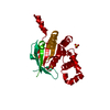
| Title | CRYSTAL STRUCTURE OF THE HELICASE DOMAIN OF THE GENE 4 PROTEIN OF BACTERIOPHAGE T7: COMPLEX WITH DATP | ||||||
 Components Components | DNA PRIMASE/HELICASE | ||||||
 Keywords Keywords |  TRANSFERASE / RECA-TYPE PROTEIN FOLD TRANSFERASE / RECA-TYPE PROTEIN FOLD | ||||||
| Function / homology |  Function and homology informationDNA primase activity / 5'-3' DNA helicase activity / viral DNA genome replication / DNA helicase activity / Transferases; Transferring phosphorus-containing groups; Nucleotidyltransferases / single-stranded DNA binding / DNA helicase / ATP hydrolysis activity / zinc ion binding / ATP binding / identical protein binding Function and homology informationDNA primase activity / 5'-3' DNA helicase activity / viral DNA genome replication / DNA helicase activity / Transferases; Transferring phosphorus-containing groups; Nucleotidyltransferases / single-stranded DNA binding / DNA helicase / ATP hydrolysis activity / zinc ion binding / ATP binding / identical protein bindingSimilarity search - Function | ||||||
| Biological species |   Enterobacteria phage T7 (virus) Enterobacteria phage T7 (virus) | ||||||
| Method | X-RAY DIFFRACTION / SYNCHROTRON / Resolution: 2.3 Å | ||||||
 Authors Authors | Sawaya, M.R. / Guo, S. / Tabor, S. / Richardson, C.C. / Ellenberger, T. | ||||||
 Citation Citation | Journal: Cell(Cambridge,Mass.) / Year: 1999 Title: Crystal structure of the helicase domain from the replicative helicase-primase of bacteriophage T7. Authors: Sawaya, M.R. / Guo, S. / Tabor, S. / Richardson, C.C. / Ellenberger, T. | ||||||
| History |
|
- Structure visualization
| Structure viewer | Molecule: MolmilJmol/JSmol |
|---|
- Downloads & links
-Download
| PDBx/mmCIF format | 1cr2.cif.gz | 62.3 KB | Display | PDBx/mmCIF format |
|---|---|---|---|---|
| PDB format | pdb1cr2.ent.gz | 44.3 KB | Display | PDB format |
| PDBx/mmJSON format | 1cr2.json.gz | Tree view | PDBx/mmJSON format | |
| Others |  Other downloads Other downloads |
-Validation report
| Arichive directory | https://data.pdbj.org/pub/pdb/validation_reports/cr/1cr2ftp://data.pdbj.org/pub/pdb/validation_reports/cr/1cr2 | HTTPS FTP |
|---|
-Related structure data


-Links
 PDBj
PDBj


- Assembly
| Deposited unit | 
| ||||||||
|---|---|---|---|---|---|---|---|---|---|
| 1 | x 6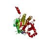
| ||||||||
| Unit cell |
| ||||||||
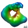
| Details | The filament may be generated by applying the cyrstallographic 6 sub 1 screw symmetry. |
-Components
| #1: Protein | Mass: 32831.977 Da / Num. of mol.: 1 / Fragment: HELICASE DOMAIN Source method: isolated from a genetically manipulated source Source: (gene. exp.) Enterobacteria phage T7 (virus) / Genus: T7-like viruses / Plasmid: PET17B / Production host:  Escherichia coli (E. coli) Escherichia coli (E. coli)References: UniProt: P03692, Transferases; Transferring phosphorus-containing groups; Nucleotidyltransferases | ||||
|---|---|---|---|---|---|
| #2: Chemical | Sulfate  Mass: 96.063 Da / Num. of mol.: 2 / Source method: obtained synthetically / Formula: SO4 Mass: 96.063 Da / Num. of mol.: 2 / Source method: obtained synthetically / Formula: SO4#3: Chemical | ChemComp-DTP / | Deoxyadenosine triphosphate  Mass: 491.182 Da / Num. of mol.: 1 / Source method: obtained synthetically / Formula: C10H16N5O12P3 Mass: 491.182 Da / Num. of mol.: 1 / Source method: obtained synthetically / Formula: C10H16N5O12P3#4: Water | ChemComp-HOH / | Water Mass: 18.015 Da / Num. of mol.: 49 / Source method: isolated from a natural source / Formula: H2O Mass: 18.015 Da / Num. of mol.: 49 / Source method: isolated from a natural source / Formula: H2O |
-Experimental details
-Experiment
| Experiment | Method: X-RAY DIFFRACTION / Number of used crystals: 1 |
|---|
- Sample preparation
| Crystal | Density Matthews: 2.42 Å3/Da / Density % sol: 49.21 % | |||||||||||||||||||||||||||||||||||
|---|---|---|---|---|---|---|---|---|---|---|---|---|---|---|---|---|---|---|---|---|---|---|---|---|---|---|---|---|---|---|---|---|---|---|---|---|
| Crystal grow | Temperature: 298 K / Method: vapor diffusion, sitting drop / pH: 9 Details: ammonium sulfate, ACES, pH 9.00, VAPOR DIFFUSION, SITTING DROP, temperature 298K | |||||||||||||||||||||||||||||||||||
| Crystal grow | *PLUS pH: 7.5 | |||||||||||||||||||||||||||||||||||
| Components of the solutions | *PLUS
|
-Data collection
| Diffraction | Mean temperature: 113 K |
|---|---|
| Diffraction source | Source: SYNCHROTRON / Site: NSLS  / Beamline: X25 / Wavelength: 1.1 / Beamline: X25 / Wavelength: 1.1 |
| Detector | Type: BRANDEIS - B4 / Detector: CCD / Date: Mar 3, 1999 |
| Radiation | Protocol: SINGLE WAVELENGTH / Monochromatic (M) / Laue (L): M / Scattering type: x-ray |
| Radiation wavelength | Wavelength: 1.1 Å / Relative weight: 1 |
| Reflection | Resolution: 2.3→20 Å / Num. all: 13072 / Num. obs: 13072 / % possible obs: 96.1 % / Observed criterion σ(F): 0 / Observed criterion σ(I): 0 / Redundancy: 11.7 % / Biso Wilson estimate: 42.4 Å2 / Rmerge(I) obs: 0.063 / Net I/σ(I): 28 |
| Reflection shell | Resolution: 2.31→2.46 Å / Redundancy: 7.6 % / Rmerge(I) obs: 0.149 / Num. unique all: 2259 / % possible all: 100 |
| Reflection | *PLUS Num. measured all: 153764 |
| Reflection shell | *PLUS % possible obs: 100 % |
- Processing
| Software |
| |||||||||||||||||||||||||
|---|---|---|---|---|---|---|---|---|---|---|---|---|---|---|---|---|---|---|---|---|---|---|---|---|---|---|
| Refinement | Resolution: 2.3→20 Å / Cross valid method: THROUGHOUT / σ(F): 0 / σ(I): 0 / Stereochemistry target values: ENGH & HUBER
| |||||||||||||||||||||||||
| Refinement step | Cycle: LAST / Resolution: 2.3→20 Å
| |||||||||||||||||||||||||
| Refine LS restraints |
| |||||||||||||||||||||||||
| Software | *PLUS Name: X-PLOR / Version: 3.851 / Classification: refinement | |||||||||||||||||||||||||
| Refinement | *PLUS Rfactor Rwork: 0.247 | |||||||||||||||||||||||||
| Solvent computation | *PLUS | |||||||||||||||||||||||||
| Displacement parameters | *PLUS |
