 Movie
Movie Controller


+ Open data

- Basic information
| Entry | Database: EMDB / ID: EMD-9011 | |||||||||
|---|---|---|---|---|---|---|---|---|---|---|
| Title | Bax with activating Bid peptide inserted into nanodisc. | |||||||||
 Map data Map data | Bax with activating Bid peptide inserted in nanodiscs | |||||||||
 Sample Sample |
| |||||||||
| Biological species |   Homo sapiens (human) Homo sapiens (human) | |||||||||
| Method | single particle reconstruction / cryo EM / Resolution: 28.0 Å | |||||||||
 Authors Authors | Volkmann N / Hanein D | |||||||||
| Funding support |  United States, 1 items United States, 1 items
| |||||||||
 Citation Citation | Journal: Structure / Year: 2019 Title: Biophysical Characterization of a Nanodisc with and without BAX: An Integrative Study Using Molecular Dynamics Simulations and Cryo-EM. Authors: Cesar A López / Mark F Swift / Xiao-Ping Xu / Dorit Hanein / Niels Volkmann / S Gnanakaran / Abstract: B cell lymphoma-2-associated X protein (BAX) plays a pivotal role in triggering cell apoptosis by permeabilizing the mitochondrial outer membrane. Contrary to previous findings, recent electron ...B cell lymphoma-2-associated X protein (BAX) plays a pivotal role in triggering cell apoptosis by permeabilizing the mitochondrial outer membrane. Contrary to previous findings, recent electron microscopy (EM) experiments showed that BAX monomers are able to perturb phospholipid nanodiscs (NDs) by forming lipidic pores. Here, we provide structural and thermodynamic interpretation of such data using multiscale resolution molecular dynamics (MD) simulations. Our results suggest that BAX is able to disrupt the stability, lateral packing and enhance the desorption propensity of the lipids in the ND, resulting in the formation of a stable toroidal-like pore. These findings prompted to re-evaluate the previously reported cryo-EM data to generate an improved reconstruction, thereby allowing for a more accurate localization of BAX in the EM map. We conclude that the reduced stability of the BAX-embedded ND eliminates the necessity of forming active BAX oligomers for its disruption. | |||||||||
| History |
|
- Structure visualization
| Movie |
 Movie viewer Movie viewer |
|---|---|
| Structure viewer | EM map: SurfViewMolmilJmol/JSmol |
| Supplemental images |

- Downloads & links
-EMDB archive
| Map data | emd_9011.map.gz | 45.4 KB | EMDB map data format | |
|---|---|---|---|---|
| Header (meta data) | emd-9011-v30.xmlemd-9011.xml | 7.7 KB 7.7 KB | Display Display | EMDB header |
| Images | 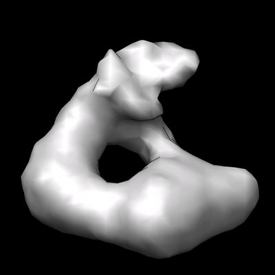 emd_9011.png emd_9011.png | 66.4 KB | ||
| Archive directory |  http://ftp.pdbj.org/pub/emdb/structures/EMD-9011ftp://ftp.pdbj.org/pub/emdb/structures/EMD-9011 http://ftp.pdbj.org/pub/emdb/structures/EMD-9011ftp://ftp.pdbj.org/pub/emdb/structures/EMD-9011 | HTTPS FTP |
-Related structure data
| Similar structure data |
|---|


-Links
| EMDB pages | EMDB (EBI/PDBe) / EMDataResource |
|---|
-Map
| File | Download / File: emd_9011.map.gz / Format: CCP4 / Size: 251 KB / Type: IMAGE STORED AS FLOATING POINT NUMBER (4 BYTES) | ||||||||||||||||||||||||||||||||||||||||||||||||||||||||||||||||||||
|---|---|---|---|---|---|---|---|---|---|---|---|---|---|---|---|---|---|---|---|---|---|---|---|---|---|---|---|---|---|---|---|---|---|---|---|---|---|---|---|---|---|---|---|---|---|---|---|---|---|---|---|---|---|---|---|---|---|---|---|---|---|---|---|---|---|---|---|---|---|
| Annotation | Bax with activating Bid peptide inserted in nanodiscs | ||||||||||||||||||||||||||||||||||||||||||||||||||||||||||||||||||||
| Voxel size | X=Y=Z: 6.76 Å | ||||||||||||||||||||||||||||||||||||||||||||||||||||||||||||||||||||
| Density |
| ||||||||||||||||||||||||||||||||||||||||||||||||||||||||||||||||||||
| Symmetry | Space group: 1 | ||||||||||||||||||||||||||||||||||||||||||||||||||||||||||||||||||||
| Details | EMDB XML:
CCP4 map header:
| ||||||||||||||||||||||||||||||||||||||||||||||||||||||||||||||||||||
-Supplemental data
- Sample components
-Entire : Complex of Bax and activating Bid peptide inserted in nanodisc
| Entire | Name: Complex of Bax and activating Bid peptide inserted in nanodisc |
|---|---|
| Components |
|
-Supramolecule #1: Complex of Bax and activating Bid peptide inserted in nanodisc
| Supramolecule | Name: Complex of Bax and activating Bid peptide inserted in nanodisc type: complex / ID: 1 / Parent: 0 |
|---|---|
| Source (natural) | Organism: Homo sapiens (human) |
| Recombinant expression | Organism:  Escherichia coli (E. coli) Escherichia coli (E. coli) |
-Experimental details
-Structure determination
| Method | cryo EM |
|---|---|
 Processing Processing | single particle reconstruction |
| Aggregation state | particle |
-Sample preparation
| Buffer | pH: 8 |
|---|---|
| Vitrification | Cryogen name: ETHANE |
- Electron microscopy
| Microscope | FEI TECNAI F20 |
|---|---|
| Electron beam | Acceleration voltage: 200 kV / Electron source: FIELD EMISSION GUN |
| Electron optics | Illumination mode: FLOOD BEAM / Imaging mode: BRIGHT FIELDBright-field microscopy |
| Image recording | Film or detector model: KODAK SO-163 FILM / Average electron dose: 25.0 e/Å2 |
| Experimental equipment |  Model: Tecnai F20 / Image courtesy: FEI Company |
-Image processing
| CTF correction | Software - Name: EMAN2 |
|---|---|
| Startup model | Type of model: OTHER Details: Previously obtained reconstructions of the same sample |
| Initial angle assignment | Type: PROJECTION MATCHING / Software - Name: EMAN2 |
| Final angle assignment | Type: PROJECTION MATCHING / Software - Name: EMAN2 |
| Final reconstruction | Resolution.type: BY AUTHOR / Resolution: 28.0 Å / Resolution method: FSC 0.5 CUT-OFF / Number images used: 4312 |
