 Movie
Movie Controller

[English] 日本語
 Yorodumi
- PDB-1a71: TERNARY COMPLEX OF AN ACTIVE SITE DOUBLE MUTANT OF HORSE LIVER AL... -
+ Open data

- Basic information
| Entry | Database: PDB / ID: 1a71 | ||||||
|---|---|---|---|---|---|---|---|
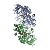
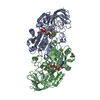
| Title | TERNARY COMPLEX OF AN ACTIVE SITE DOUBLE MUTANT OF HORSE LIVER ALCOHOL DEHYDROGENASE, PHE93=>TRP, VAL203=>ALA WITH NAD AND TRIFLUOROETHANOL | ||||||
 Components Components | LIVER ALCOHOL DEHYDROGENASE Alcohol dehydrogenase Alcohol dehydrogenase | ||||||
 Keywords Keywords | OXIDOREDUCTASE / OXIDOREDUCTASE (NAD(A)-CHOH(D)) / LIVER / ALCOHOL / DEHYDROGENASE / LADH / ACTIVE SITE MUTANT | ||||||
| Function / homology |  Function and homology information Function and homology informationalcohol dehydrogenase (NAD+) activity, zinc-dependent / : / all-trans-retinol dehydrogenase (NAD+) activity / alcohol dehydrogenase / retinol metabolic process / retinoic acid metabolic process / zinc ion binding / cytosolSimilarity search - Function | ||||||
| Biological species |  Equus caballus (horse) Equus caballus (horse) | ||||||
| Method | X-RAY DIFFRACTION / MOLECULAR REPLACEMENT / Resolution: 2 Å | ||||||
 Authors Authors | Colby, T.D. / Bahnson, B.J. / Chin, J.K. / Klinman, J.P. / Goldstein, B.M. | ||||||
 Citation Citation | Journal: Biochemistry / Year: 1998 Title: Active site modifications in a double mutant of liver alcohol dehydrogenase: structural studies of two enzyme-ligand complexes. Authors: Colby, T.D. / Bahnson, B.J. / Chin, J.K. / Klinman, J.P. / Goldstein, B.M. #1: Journal: Proc.Natl.Acad.Sci.USA / Year: 1997Title: A Link between Protein Structure and Enzyme Catalyzed Hydrogen Tunneling Authors: Bahnson, B.J. / Colby, T.D. / Chin, J.K. / Goldstein, B.M. / Klinman, J.P. | ||||||
| History |
|
- Structure visualization
| Structure viewer | Molecule: MolmilJmol/JSmol |
|---|
- Downloads & links
-Download
| PDBx/mmCIF format | 1a71.cif.gz | 154.9 KB | Display | PDBx/mmCIF format |
|---|---|---|---|---|
| PDB format | pdb1a71.ent.gz | 121.4 KB | Display | PDB format |
| PDBx/mmJSON format | 1a71.json.gz | Tree view | PDBx/mmJSON format | |
| Others |  Other downloads Other downloads |
-Validation report
| Arichive directory | https://data.pdbj.org/pub/pdb/validation_reports/a7/1a71ftp://data.pdbj.org/pub/pdb/validation_reports/a7/1a71 | HTTPS FTP |
|---|
-Related structure data
| Related structure data | 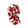 1a72C 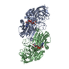 2ohxS S: Starting model for refinement C: citing same article ( |
|---|---|
| Similar structure data |

-Links
 PDBj
PDBj

- Assembly
| Deposited unit | 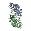
| ||||||||
|---|---|---|---|---|---|---|---|---|---|
| 1 |
| ||||||||
| Unit cell |
|
-Components
| #1: Protein | Alcohol dehydrogenase Mass: 39864.258 Da / Num. of mol.: 2 / Mutation: F93W, V203A Source method: isolated from a genetically manipulated source Source: (gene. exp.) Equus caballus (horse) / Cellular location: CYTOPLASM / Gene: LADH F93W V203A / Organ: LIVER / Plasmid: PHAGEMID PBPP-LADH / Cellular location (production host): CYTOPLASM / Gene (production host): LADH F93W,V203A / Production host:  Escherichia coli (E. coli) / Strain (production host): XL1-BLUE / References: UniProt: P00327, alcohol dehydrogenase Escherichia coli (E. coli) / Strain (production host): XL1-BLUE / References: UniProt: P00327, alcohol dehydrogenase#2: Chemical | ChemComp-ZN /   Mass: 65.409 Da / Num. of mol.: 4 / Source method: obtained synthetically / Formula: Zn Mass: 65.409 Da / Num. of mol.: 4 / Source method: obtained synthetically / Formula: Zn#3: Chemical | Nicotinamide adenine dinucleotide  Mass: 663.425 Da / Num. of mol.: 2 / Source method: obtained synthetically / Formula: C21H27N7O14P2 / Comment: NAD*YM Mass: 663.425 Da / Num. of mol.: 2 / Source method: obtained synthetically / Formula: C21H27N7O14P2 / Comment: NAD*YM#4: Chemical | 2,2,2-Trifluoroethanol  Mass: 100.040 Da / Num. of mol.: 2 / Source method: obtained synthetically / Formula: C2H3F3O Mass: 100.040 Da / Num. of mol.: 2 / Source method: obtained synthetically / Formula: C2H3F3O#5: Water | ChemComp-HOH / | Water Mass: 18.015 Da / Num. of mol.: 179 / Source method: isolated from a natural source / Formula: H2O Mass: 18.015 Da / Num. of mol.: 179 / Source method: isolated from a natural source / Formula: H2O |
|---|
-Experimental details
-Experiment
| Experiment | Method: X-RAY DIFFRACTION / Number of used crystals: 1 |
|---|
- Sample preparation
| Crystal | Density Matthews: 2.3 Å3/Da / Density % sol: 47.99 % | |||||||||||||||||||||||||||||||||||
|---|---|---|---|---|---|---|---|---|---|---|---|---|---|---|---|---|---|---|---|---|---|---|---|---|---|---|---|---|---|---|---|---|---|---|---|---|
| Crystal grow | Method: vapor diffusion, hanging drop / pH: 8.4 Details: 4MICROLITER HANGING DROPS TRIS PH 8.4 AT 4C, 5MM TRIFLUROETHANOL, 4% PEG400 EQUILIBRATED AGAINST WELLS CONTAINING 5MM TFE AND 18%PEG 400, vapor diffusion - hanging drop | |||||||||||||||||||||||||||||||||||
| Crystal grow | *PLUS Temperature: 4 ℃ / Method: vapor diffusion | |||||||||||||||||||||||||||||||||||
| Components of the solutions | *PLUS
|
-Data collection
| Diffraction | Mean temperature: 100 K |
|---|---|
| Diffraction source | Source: ROTATING ANODE / Type: RIGAKU RUH2R / Wavelength: 1.5418 |
| Detector | Type: RIGAKU RAXIS II / Detector: IMAGE PLATE / Date: May 1, 1996 / Details: YALE MIRRORS |
| Radiation | Monochromatic (M) / Laue (L): M / Scattering type: x-ray |
| Radiation wavelength | Wavelength: 1.5418 Å / Relative weight: 1 |
| Reflection | Resolution: 2→50 Å / Num. obs: 38046 / % possible obs: 77.1 % / Observed criterion σ(I): 2 / Redundancy: 1.7 % / Biso Wilson estimate: 21 Å2 / Rmerge(I) obs: 0.071 / Net I/σ(I): 7.5 |
| Reflection shell | Resolution: 2→2.03 Å / Redundancy: 1.7 % / Rmerge(I) obs: 0.34 / Mean I/σ(I) obs: 2 / % possible all: 42.3 |
| Reflection | *PLUS Num. measured all: 77199 |
| Reflection shell | *PLUS % possible obs: 42.3 % |
- Processing
| Software |
| ||||||||||||||||||||||||||||||||||||||||||||||||||||||||||||||||||||||||||||||||
|---|---|---|---|---|---|---|---|---|---|---|---|---|---|---|---|---|---|---|---|---|---|---|---|---|---|---|---|---|---|---|---|---|---|---|---|---|---|---|---|---|---|---|---|---|---|---|---|---|---|---|---|---|---|---|---|---|---|---|---|---|---|---|---|---|---|---|---|---|---|---|---|---|---|---|---|---|---|---|---|---|---|
| Refinement | Method to determine structure: MOLECULAR REPLACEMENT Starting model: PDB ENTRY 2OHX Resolution: 2→10 Å / Rfactor Rfree error: 0.005 / Data cutoff high absF: 1000000 / Data cutoff low absF: 0.1 / Isotropic thermal model: RESTRAINED Cross valid method: UP TO LAST CG MINIMIZATION THROUGHOUT UNTIL LAST CG REFINEMENT σ(F): 2
| ||||||||||||||||||||||||||||||||||||||||||||||||||||||||||||||||||||||||||||||||
| Displacement parameters | Biso mean: 22.1 Å2
| ||||||||||||||||||||||||||||||||||||||||||||||||||||||||||||||||||||||||||||||||
| Refine analyze | Luzzati coordinate error obs: 0.39 Å / Luzzati d res low obs: 5 Å | ||||||||||||||||||||||||||||||||||||||||||||||||||||||||||||||||||||||||||||||||
| Refinement step | Cycle: LAST / Resolution: 2→10 Å
| ||||||||||||||||||||||||||||||||||||||||||||||||||||||||||||||||||||||||||||||||
| Refine LS restraints |
| ||||||||||||||||||||||||||||||||||||||||||||||||||||||||||||||||||||||||||||||||
| LS refinement shell | Resolution: 2→2.09 Å / Rfactor Rfree error: 0.02 / Total num. of bins used: 8
| ||||||||||||||||||||||||||||||||||||||||||||||||||||||||||||||||||||||||||||||||
| Xplor file |
| ||||||||||||||||||||||||||||||||||||||||||||||||||||||||||||||||||||||||||||||||
| Software | *PLUS Name: X-PLOR / Version: 3.1 / Classification: refinement | ||||||||||||||||||||||||||||||||||||||||||||||||||||||||||||||||||||||||||||||||
| Refinement | *PLUS | ||||||||||||||||||||||||||||||||||||||||||||||||||||||||||||||||||||||||||||||||
| Solvent computation | *PLUS | ||||||||||||||||||||||||||||||||||||||||||||||||||||||||||||||||||||||||||||||||
| Displacement parameters | *PLUS | ||||||||||||||||||||||||||||||||||||||||||||||||||||||||||||||||||||||||||||||||
| Refine LS restraints | *PLUS
| ||||||||||||||||||||||||||||||||||||||||||||||||||||||||||||||||||||||||||||||||
| LS refinement shell | *PLUS Rfactor obs: 0.268 |
