 Movie
Movie Controller

[English] 日本語
 Yorodumi
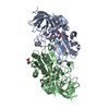
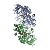
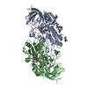
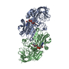
- PDB-3oq6: Horse liver alcohol dehydrogenase A317C mutant complexed with NAD... -
+ Open data

- Basic information
| Entry | Database: PDB / ID: 3oq6 | ||||||
|---|---|---|---|---|---|---|---|
| Title | Horse liver alcohol dehydrogenase A317C mutant complexed with NAD+ and 2,3,4,5,6-pentafluorobenzyl alcohol | ||||||
 Components Components | Alcohol dehydrogenase E chain | ||||||
 Keywords Keywords |  OXIDOREDUCTASE / Rossmann fold / alcohol metabolism / NAD OXIDOREDUCTASE / Rossmann fold / alcohol metabolism / NAD | ||||||
| Function / homology |  Function and homology information Function and homology informationalcohol dehydrogenase (NAD+) activity, zinc-dependent / : / all-trans-retinol dehydrogenase (NAD+) activity / alcohol dehydrogenase / retinol metabolic process / retinoic acid metabolic process / zinc ion binding / cytosolSimilarity search - Function | ||||||
| Biological species |  Equus caballus (horse) Equus caballus (horse) | ||||||
| Method | X-RAY DIFFRACTION / SYNCHROTRON / MOLECULAR REPLACEMENT / Resolution: 1.2 Å | ||||||
 Authors Authors | Plapp, B.V. / Herdendorf, T.J. | ||||||
 Citation Citation | Journal: Chem.Biol.Interact / Year: 2011 Title: Origins of the high catalytic activity of human alcohol dehydrogenase 4 studied with horse liver A317C alcohol dehydrogenase. Authors: Herdendorf, T.J. / Plapp, B.V. #1: Journal: Biochemistry / Year: 1994Title: Structures of horse liver alcohol dehydrogenase complexed with NAD and substituted benzyl alcohols Authors: Ramaswamy, S. / Eklund, H. / Plapp, B.V. | ||||||
| History |
|
- Structure visualization
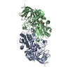
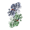
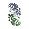
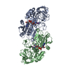
| Structure viewer | Molecule: MolmilJmol/JSmol |
|---|
- Downloads & links
-Download
| PDBx/mmCIF format | 3oq6.cif.gz | 348.6 KB | Display | PDBx/mmCIF format |
|---|---|---|---|---|
| PDB format | pdb3oq6.ent.gz | 283.2 KB | Display | PDB format |
| PDBx/mmJSON format | 3oq6.json.gz | Tree view | PDBx/mmJSON format | |
| Others |  Other downloads Other downloads |
-Validation report
| Arichive directory | https://data.pdbj.org/pub/pdb/validation_reports/oq/3oq6ftp://data.pdbj.org/pub/pdb/validation_reports/oq/3oq6 | HTTPS FTP |
|---|
-Related structure data
| Related structure data |  1hldS S: Starting model for refinement |
|---|---|
| Similar structure data |


-Links
 PDBj
PDBj

- Assembly
| Deposited unit | 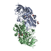
| ||||||||
|---|---|---|---|---|---|---|---|---|---|
| 1 |
| ||||||||
| Unit cell |
|
-Components
-Protein , 1 types, 2 molecules AB
| #1: Protein | Mass: 39885.336 Da / Num. of mol.: 2 / Mutation: A317C Source method: isolated from a genetically manipulated source Source: (gene. exp.) Equus caballus (horse) / Gene: ADH E / Plasmid: pET23d(+) / Production host:  Escherichia coli (E. coli) / Strain (production host): BL21(lamdaDE3) / References: UniProt: P00327, alcohol dehydrogenase Escherichia coli (E. coli) / Strain (production host): BL21(lamdaDE3) / References: UniProt: P00327, alcohol dehydrogenase |
|---|
-Non-polymers , 5 types, 1109 molecules 
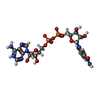
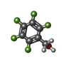


| #2: Chemical | ChemComp-ZN /  Mass: 65.409 Da / Num. of mol.: 4 / Source method: obtained synthetically / Formula: Zn Mass: 65.409 Da / Num. of mol.: 4 / Source method: obtained synthetically / Formula: Zn#3: Chemical | Nicotinamide adenine dinucleotide Mass: 663.425 Da / Num. of mol.: 2 / Source method: obtained synthetically / Formula: C21H27N7O14P2 Mass: 663.425 Da / Num. of mol.: 2 / Source method: obtained synthetically / Formula: C21H27N7O14P2#4: Chemical |  Mass: 198.090 Da / Num. of mol.: 2 / Source method: obtained synthetically / Formula: C7H3F5O Mass: 198.090 Da / Num. of mol.: 2 / Source method: obtained synthetically / Formula: C7H3F5O#5: Chemical | 2-Methyl-2,4-pentanediol Mass: 118.174 Da / Num. of mol.: 3 / Source method: obtained synthetically / Formula: C6H14O2 / Comment: precipitant*YM Mass: 118.174 Da / Num. of mol.: 3 / Source method: obtained synthetically / Formula: C6H14O2 / Comment: precipitant*YM#6: Water | ChemComp-HOH / | WaterMass: 18.015 Da / Num. of mol.: 1098 / Source method: isolated from a natural source / Formula: H2O |
|---|
-Experimental details
-Experiment
| Experiment | Method: X-RAY DIFFRACTION / Number of used crystals: 1 |
|---|
- Sample preparation
| Crystal | Density Matthews: 2.39 Å3/Da / Density % sol: 48.51 % |
|---|---|
| Crystal grow | Temperature: 298 K / Method: microdialysis / pH: 7 Details: 50 mM ammonium TES [N-tris(hydroxymethyl)-2-aminoethane sulfonate], 25% MRD, 11 mg/ml protein, 11 mM NAD+, 5 mM 2,3,4,5,6-pentafluorobenzyl alcohol, pH 7.0, MICRODIALYSIS, temperature 298K |
-Data collection
| Diffraction | Mean temperature: 100 K | ||||||||||||||||||||||||||||||||||||||||||||||||||||||||||||||||||||||||||||||||||||||||
|---|---|---|---|---|---|---|---|---|---|---|---|---|---|---|---|---|---|---|---|---|---|---|---|---|---|---|---|---|---|---|---|---|---|---|---|---|---|---|---|---|---|---|---|---|---|---|---|---|---|---|---|---|---|---|---|---|---|---|---|---|---|---|---|---|---|---|---|---|---|---|---|---|---|---|---|---|---|---|---|---|---|---|---|---|---|---|---|---|---|
| Diffraction source | Source: SYNCHROTRON / Site: ALS  / Beamline: 4.2.2 / Wavelength: 0.8 Å / Beamline: 4.2.2 / Wavelength: 0.8 Å | ||||||||||||||||||||||||||||||||||||||||||||||||||||||||||||||||||||||||||||||||||||||||
| Detector | Type: NOIR-1 / Detector: CCD / Date: Mar 27, 2009 / Details: SAGGITALLY FOCUSED MIRRORS | ||||||||||||||||||||||||||||||||||||||||||||||||||||||||||||||||||||||||||||||||||||||||
| Radiation | Protocol: SINGLE WAVELENGTH / Monochromatic (M) / Laue (L): M / Scattering type: x-ray | ||||||||||||||||||||||||||||||||||||||||||||||||||||||||||||||||||||||||||||||||||||||||
| Radiation wavelength | Wavelength: 0.8 Å / Relative weight: 1 | ||||||||||||||||||||||||||||||||||||||||||||||||||||||||||||||||||||||||||||||||||||||||
| Reflection | Resolution: 1.2→90 Å / Num. obs: 214213 / % possible obs: 92.2 % / Redundancy: 3.57 % / Biso Wilson estimate: 11.5 Å2 / Rmerge(I) obs: 0.061 / Χ2: 1 / Net I/σ(I): 9.4 / Scaling rejects: 5787 | ||||||||||||||||||||||||||||||||||||||||||||||||||||||||||||||||||||||||||||||||||||||||
| Reflection shell | Diffraction-ID: 1
|
- Processing
| Software |
| ||||||||||||||||||||||||||||||||||||||||||||||||||||||||||||||||||||||||||||||||||||||||||
|---|---|---|---|---|---|---|---|---|---|---|---|---|---|---|---|---|---|---|---|---|---|---|---|---|---|---|---|---|---|---|---|---|---|---|---|---|---|---|---|---|---|---|---|---|---|---|---|---|---|---|---|---|---|---|---|---|---|---|---|---|---|---|---|---|---|---|---|---|---|---|---|---|---|---|---|---|---|---|---|---|---|---|---|---|---|---|---|---|---|---|---|
| Refinement | Method to determine structure: MOLECULAR REPLACEMENT Starting model: PDB entry 1hld Resolution: 1.2→19.85 Å / Cor.coef. Fo:Fc: 0.982 / Cor.coef. Fo:Fc free: 0.977 / WRfactor Rfree: 0.1748 / WRfactor Rwork: 0.1442 / Occupancy max: 1 / Occupancy min: 0.03 / FOM work R set: 0.8668 / SU B: 1.303 / SU ML: 0.026 / SU R Cruickshank DPI: 0.0359 / SU Rfree: 0.0362 / Cross valid method: THROUGHOUT / σ(F): 0 / ESU R Free: 0.036 / Stereochemistry target values: MAXIMUM LIKELIHOOD Details: HYDROGENS HAVE BEEN ADDED IN THE RIDING POSITIONS U VALUES: REFINED INDIVIDUALLY. B107 GLU O TOO CLOSE TO B741 O, BECAUSE OF A WOBBLE IN THE PEPTIDE BACKBONE THAT THE AUTHORS ARE UNABLE TO FIT PROPERLY
| ||||||||||||||||||||||||||||||||||||||||||||||||||||||||||||||||||||||||||||||||||||||||||
| Solvent computation | Ion probe radii: 0.8 Å / Shrinkage radii: 0.8 Å / VDW probe radii: 1.4 Å / Solvent model: MASK | ||||||||||||||||||||||||||||||||||||||||||||||||||||||||||||||||||||||||||||||||||||||||||
| Displacement parameters | Biso max: 97.31 Å2 / Biso mean: 17.5778 Å2 / Biso min: 5.72 Å2
| ||||||||||||||||||||||||||||||||||||||||||||||||||||||||||||||||||||||||||||||||||||||||||
| Refinement step | Cycle: LAST / Resolution: 1.2→19.85 Å
| ||||||||||||||||||||||||||||||||||||||||||||||||||||||||||||||||||||||||||||||||||||||||||
| Refine LS restraints |
| ||||||||||||||||||||||||||||||||||||||||||||||||||||||||||||||||||||||||||||||||||||||||||
| LS refinement shell | Resolution: 1.2→1.229 Å / Total num. of bins used: 20
|
