RNA secondary structure unwinding / positive regulation of cytoplasmic translation / negative regulation of cytoplasmic translational initiation / mRNA base-pairing translational repressor activity / ornithine decarboxylase inhibitor activity / transcription antitermination factor activity, RNA binding / misfolded RNA binding / Group I intron splicing / RNA folding / positive regulation of ribosome biogenesis ...RNA secondary structure unwinding / positive regulation of cytoplasmic translation / negative regulation of cytoplasmic translational initiation / mRNA base-pairing translational repressor activity / ornithine decarboxylase inhibitor activity / transcription antitermination factor activity, RNA binding / misfolded RNA binding / Group I intron splicing / RNA folding / positive regulation of ribosome biogenesis / negative regulation of cytoplasmic translation / four-way junction DNA binding / DnaA-L2 complex / negative regulation of translational initiation / translational initiation / negative regulation of DNA-templated DNA replication initiation / regulation of mRNA stability / mRNA regulatory element binding translation repressor activity / assembly of large subunit precursor of preribosome / positive regulation of RNA splicing / transcription elongation factor complex / cytosolic ribosome assembly / DNA endonuclease activity / regulation of DNA-templated transcription elongation / transcription antitermination / regulation of cell growth / DNA-templated transcription termination / maintenance of translational fidelity / ribosomal large subunit assembly / mRNA 5'-UTR binding / ribosomal small subunit biogenesis / ribosomal small subunit assembly / small ribosomal subunit rRNA binding / リボソーム生合成 / ribosome binding / regulation of translation / 5S rRNA binding / large ribosomal subunit rRNA binding / small ribosomal subunit / cytosolic small ribosomal subunit / transferase activity / cytosolic large ribosomal subunit / cytoplasmic translation / tRNA binding / molecular adaptor activity / single-stranded RNA binding / negative regulation of translation / rRNA binding / リボソーム / structural constituent of ribosome / 翻訳 (生物学) / response to antibiotic / mRNA binding / RNA binding / zinc ion binding / 生体膜 / 細胞質基質 / 細胞質 類似検索 - 分子機能
Ribosomal protein S1 / Ribosomal protein S1-like / Ribosomal protein L1, bacterial-type / S1 domain profile. / Ribosomal protein L1, conserved site / Ribosomal protein L1 signature. / Ribosomal protein L1 / Ribosomal protein S21, conserved site / Ribosomal protein S21 signature. / Ribosomal protein L1, 3-layer alpha/beta-sandwich ...Ribosomal protein S1 / Ribosomal protein S1-like / Ribosomal protein L1, bacterial-type / S1 domain profile. / Ribosomal protein L1, conserved site / Ribosomal protein L1 signature. / Ribosomal protein L1 / Ribosomal protein S21, conserved site / Ribosomal protein S21 signature. / Ribosomal protein L1, 3-layer alpha/beta-sandwich / Ribosomal protein S14, bacterial/plastid / Ribosomal protein S1-like RNA-binding domain / Ribosomal protein L1-like / Ribosomal protein L1/ribosomal biogenesis protein / Ribosomal protein L1p/L10e family / S1 RNA binding domain / Ribosomal protein S21 superfamily / Ribosomal protein L31 type A / S1 domain / Ribosomal protein S21 / Ribosomal protein S16, conserved site / Ribosomal protein S16 signature. / Ribosomal protein S21 / Ribosomal protein L31 signature. / Ribosomal protein L31 / Ribosomal protein L31 superfamily / Ribosomal protein L31 / Ribosomal protein L16 signature 1. / Ribosomal protein L16, conserved site / Ribosomal protein L16 signature 2. / Ribosomal protein L9 signature. / Ribosomal protein L9, bacteria/chloroplast / Ribosomal protein L9, C-terminal / Ribosomal protein L9, C-terminal domain / Ribosomal protein L9, C-terminal domain superfamily / Ribosomal protein L28/L24 superfamily / Ribosomal protein L9, N-terminal domain superfamily / Ribosomal protein L9 / Ribosomal protein L9, N-terminal / Ribosomal protein L9, N-terminal domain / Ribosomal protein L28 / Ribosomal protein L35, conserved site / Ribosomal protein L35 signature. / Ribosomal protein L33, conserved site / Ribosomal protein L33 signature. / Ribosomal protein L35, non-mitochondrial / Ribosomal protein L5, bacterial-type / Ribosomal protein L18, bacterial-type / Ribosomal protein L9/RNase H1, N-terminal / Ribosomal protein S3, bacterial-type / Ribosomal protein S6, conserved site / Ribosomal protein S6 signature. / Ribosomal protein S19, bacterial-type / Ribosomal protein S7, bacterial/organellar-type / Ribosomal protein S11, bacterial-type / Ribosomal protein L27, conserved site / Ribosomal protein S13, bacterial-type / Ribosomal protein L27 signature. / Ribosomal protein S20 / Ribosomal protein S20 superfamily / Ribosomal protein S20 / Ribosomal protein S9, bacterial/plastid / Ribosomal protein S4, bacterial-type / 30S ribosomal protein S17 / Ribosomal protein S5, bacterial-type / Ribosomal protein L34, conserved site / Ribosomal protein L34 signature. / Ribosomal protein S6, plastid/chloroplast / Ribosomal protein L35 / Ribosomal protein L35 superfamily / Ribosomal protein L2, bacterial/organellar-type / Ribosomal protein S2, bacteria/mitochondria/plastid / Ribosomal protein L35 / Ribosomal L28 family / Ribosomal protein L33 / Ribosomal protein L33 / Ribosomal protein L28/L24 / Ribosomal protein L33 superfamily / Ribosomal protein L30, bacterial-type / Ribosomal protein L16 / Ribosomal protein L18 / Ribosomal L18 of archaea, bacteria, mitoch. and chloroplast / Ribosomal protein S18, conserved site / Ribosomal protein S18 signature. / L28p-like / Ribosomal protein S16 / Ribosomal protein S16 / Ribosomal protein S16 domain superfamily / Ribosomal protein S15, bacterial-type / Ribosomal protein L27 / Ribosomal L27 protein / Ribosomal protein S6 / Ribosomal protein S6 / Ribosomal protein S2 signature 2. / Ribosomal protein S6 superfamily / Ribosomal protein S12, bacterial-type / Ribosomal protein L34 / Ribosomal protein L34 / Translation elongation factor EF1B/ribosomal protein S6 / Ribosomal protein S18 類似検索 - ドメイン・相同性
Small ribosomal subunit protein bS6 / Small ribosomal subunit protein uS7 / Large ribosomal subunit protein uL15 / Large ribosomal subunit protein uL1 / Large ribosomal subunit protein bL27 / Large ribosomal subunit protein bL28 / Large ribosomal subunit protein bL31 / Large ribosomal subunit protein bL33 / Large ribosomal subunit protein bL34 / Large ribosomal subunit protein bL35 ...Small ribosomal subunit protein bS6 / Small ribosomal subunit protein uS7 / Large ribosomal subunit protein uL15 / Large ribosomal subunit protein uL1 / Large ribosomal subunit protein bL27 / Large ribosomal subunit protein bL28 / Large ribosomal subunit protein bL31 / Large ribosomal subunit protein bL33 / Large ribosomal subunit protein bL34 / Large ribosomal subunit protein bL35 / Large ribosomal subunit protein bL9 / Small ribosomal subunit protein uS10 / Small ribosomal subunit protein uS11 / Small ribosomal subunit protein uS12 / Small ribosomal subunit protein uS13 / Small ribosomal subunit protein bS16 / Small ribosomal subunit protein bS18 / Small ribosomal subunit protein uS19 / Small ribosomal subunit protein bS20 / Small ribosomal subunit protein uS2 / Small ribosomal subunit protein uS3 / Small ribosomal subunit protein uS4 / Small ribosomal subunit protein uS5 / Small ribosomal subunit protein uS8 / Small ribosomal subunit protein uS9 / Large ribosomal subunit protein uL16 / Small ribosomal subunit protein uS15 / Large ribosomal subunit protein uL30 / Small ribosomal subunit protein uS14 / Small ribosomal subunit protein uS17 / Small ribosomal subunit protein bS1 / Large ribosomal subunit protein uL18 / Large ribosomal subunit protein uL2 / Large ribosomal subunit protein uL5 / Small ribosomal subunit protein bS21 類似検索 - 構成要素
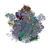
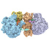
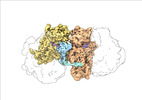
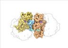
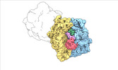
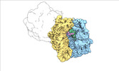
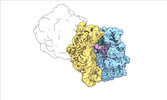
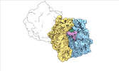
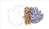
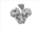
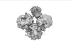
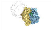
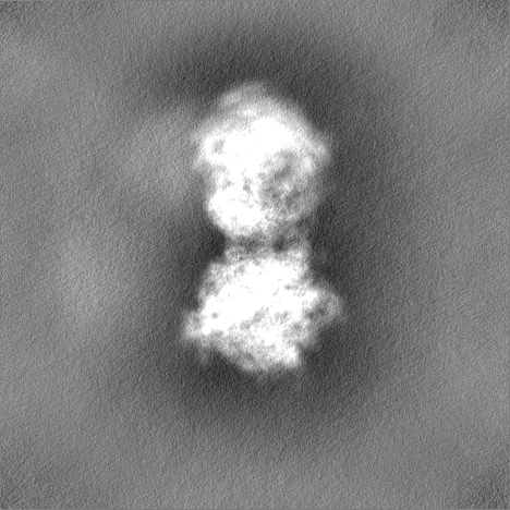
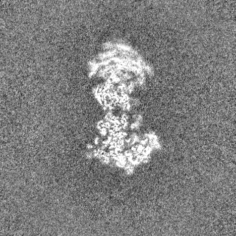
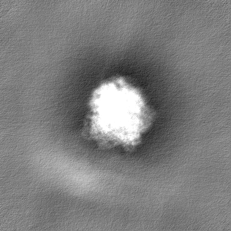
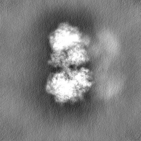
ジャーナル: Nat Commun / 年: 2024 タイトル: Transient disome complex formation in native polysomes during ongoing protein synthesis captured by cryo-EM. 著者: Timo Flügel / Magdalena Schacherl / Anett Unbehaun / Birgit Schroeer / Marylena Dabrowski / Jörg Bürger / Thorsten Mielke / Thiemo Sprink / Christoph A Diebolder / Yollete V Guillén ...著者: Timo Flügel / Magdalena Schacherl / Anett Unbehaun / Birgit Schroeer / Marylena Dabrowski / Jörg Bürger / Thorsten Mielke / Thiemo Sprink / Christoph A Diebolder / Yollete V Guillén Schlippe / Christian M T Spahn / 要旨: Structural studies of translating ribosomes traditionally rely on in vitro assembly and stalling of ribosomes in defined states. To comprehensively visualize bacterial translation, we reactivated ex ...Structural studies of translating ribosomes traditionally rely on in vitro assembly and stalling of ribosomes in defined states. To comprehensively visualize bacterial translation, we reactivated ex vivo-derived E. coli polysomes in the PURE in vitro translation system and analyzed the actively elongating polysomes by cryo-EM. We find that 31% of 70S ribosomes assemble into disome complexes that represent eight distinct functional states including decoding and termination intermediates, and a pre-nucleophilic attack state. The functional diversity of disome complexes together with RNase digest experiments suggests that paused disome complexes transiently form during ongoing elongation. Structural analysis revealed five disome interfaces between leading and queueing ribosomes that undergo rearrangements as the leading ribosome traverses through the elongation cycle. Our findings reveal at the molecular level how bL9's CTD obstructs the factor binding site of queueing ribosomes to thwart harmful collisions and illustrate how translation dynamics reshape inter-ribosomal contacts.
凍結剤: ETHANE / チャンバー内湿度: 75 % / チャンバー内温度: 277 K / 装置: FEI VITROBOT MARK IV 詳細: Withdrawn samples were spotted directly onto freshly glow-discharged holey carbon grids, blotted for 1-2 s, and flash frozen in liquid ethane using a Vitrobot Mark IV plunger (ThermoFisher ...詳細: Withdrawn samples were spotted directly onto freshly glow-discharged holey carbon grids, blotted for 1-2 s, and flash frozen in liquid ethane using a Vitrobot Mark IV plunger (ThermoFisher Scientific) after a wait time of 40 s at 4 degrees Celcius..
詳細
In vitro translation reactions were performed in the PURE translation system using the PURExpress delta ribosome kit (NEB, #E3313S). Translation reactions were supplemented with 0.8 U/uL RNAsin Plus RNase Inhibitor (Promega, N261B). SolA, factor mix, and RNAsin Plus were combined on ice, followed by a preincubation at 37 degrees Celcius for 2 min, and added directly to polysomes (700 nM final concentration) that had been preincubated at 37 degrees Celsius for 2 min. After 1 min reaction time, 4 uL of the reaction mixture were withdrawn for plunge freezing.
Chain - Source name: PDB / Chain - Initial model type: experimental model
ソフトウェア
名称: Coot (ver. 0.9.6)
詳細
The atomic model of the E. coli 70S ribosome (PDB ID: 7N1P) was initially docked into the postprocessed density maps with UCSF Chimera and manually adjusted in Coot. All models were refined over multiple rounds using the module phenix.real_space_refine in PHENIX and interactive model building and refinement in Coot, using libG restraints for the RNAs.
精密化
空間: REAL / プロトコル: RIGID BODY FIT 当てはまり具合の基準: Cross-correlation coefficient
 ムービー
ムービー コントローラー


 データを開く
 基本情報
基本情報
 マップデータ
 試料
試料 キーワード
キーワード polysome (ポリソーム) /
  機能・相同性情報
 データ登録者
 ドイツ, 1件
  引用
引用 構造の表示
構造の表示
 ダウンロードとリンク
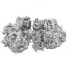
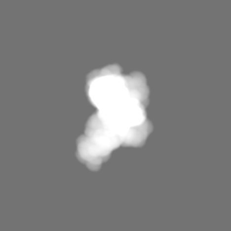
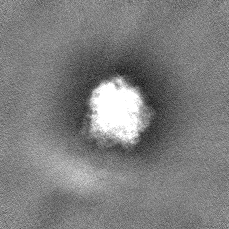
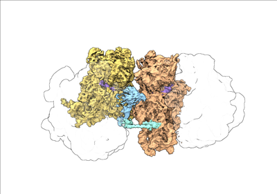 emd_19054.png
 http://ftp.pdbj.org/pub/emdb/structures/EMD-19054


 Z
Z Y
Y X
X


 試料の構成要素


 解析
解析 電子顕微鏡法