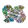
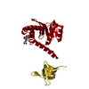
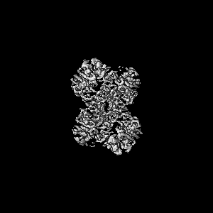
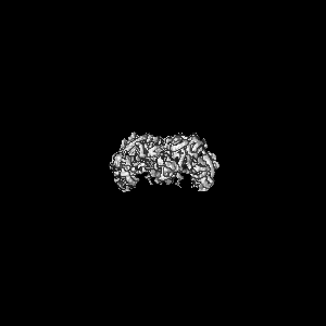
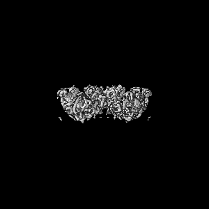
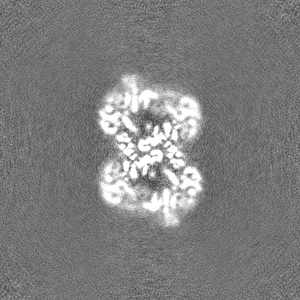
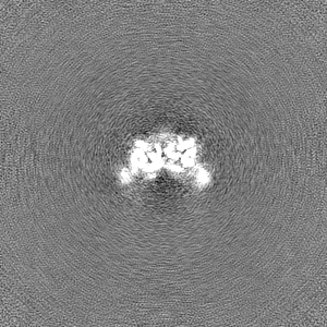
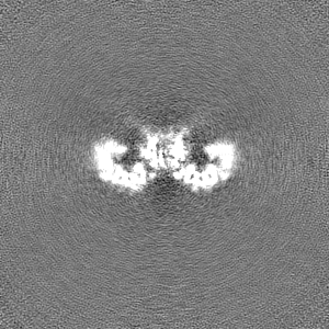
ジャーナル: Science / 年: 2017 タイトル: A supramolecular assembly mediates lentiviral DNA integration. 著者: Allison Ballandras-Colas / Daniel P Maskell / Erik Serrao / Julia Locke / Paolo Swuec / Stefán R Jónsson / Abhay Kotecha / Nicola J Cook / Valerie E Pye / Ian A Taylor / Valgerdur ...著者: Allison Ballandras-Colas / Daniel P Maskell / Erik Serrao / Julia Locke / Paolo Swuec / Stefán R Jónsson / Abhay Kotecha / Nicola J Cook / Valerie E Pye / Ian A Taylor / Valgerdur Andrésdóttir / Alan N Engelman / Alessandro Costa / Peter Cherepanov / 要旨: Retroviral integrase (IN) functions within the intasome nucleoprotein complex to catalyze insertion of viral DNA into cellular chromatin. Using cryo-electron microscopy, we now visualize the ...Retroviral integrase (IN) functions within the intasome nucleoprotein complex to catalyze insertion of viral DNA into cellular chromatin. Using cryo-electron microscopy, we now visualize the functional maedi-visna lentivirus intasome at 4.9 angstrom resolution. The intasome comprises a homo-hexadecamer of IN with a tetramer-of-tetramers architecture featuring eight structurally distinct types of IN protomers supporting two catalytically competent subunits. The conserved intasomal core, previously observed in simpler retroviral systems, is formed between two IN tetramers, with a pair of C-terminal domains from flanking tetramers completing the synaptic interface. Our results explain how HIV-1 IN, which self-associates into higher-order multimers, can form a functional intasome, reconcile the bulk of early HIV-1 IN biochemical and structural data, and provide a lentiviral platform for design of HIV-1 IN inhibitors.
凍結剤: ETHANE / チャンバー内湿度: 100 % / チャンバー内温度: 293.15 K / 装置: FEI VITROBOT MARK IV 詳細: To lower salt concentration before plunge-freezing, the grids were blotted for 0.5 s, immediately hydrated with a 4-ul drop of 200 mM NaCl, 3 mM CaCl2 and 25 mM BisTris-HCl pH 6.5 and blotted ...詳細: To lower salt concentration before plunge-freezing, the grids were blotted for 0.5 s, immediately hydrated with a 4-ul drop of 200 mM NaCl, 3 mM CaCl2 and 25 mM BisTris-HCl pH 6.5 and blotted again for 2.5 s followed by plunging into liquid ethane..
詳細
A 4 ul drop of freshly prepared intasome in 1 M NaCl, 3 mM CaCl2 and 25 mM BisTris-HCl pH 6.5 was applied onto glow-discharged lacey carbon grids coated with ultrathin carbon (product 01824, Ted Pella). The grids were incubated for 30 s under 100% humidity in a Vitrobot Mark IV (FEI) at 20 oC. To lower salt concentration before plunge-freezing, the grids were blotted for 0.5 s, immediately hydrated with a 4 ul drop of 200 mM NaCl, 3 mM CaCl2, and 25 mM BisTris-HCl pH 6.5 and blotted again for 2.5 s, followed by plunging into liquid ethane.
5M0Q model was docked to new map (updated relion version and pixel size corrected) in Chimera and refined using phenix.real_space refine and interactively adjusted in coot.
精密化
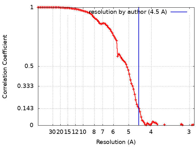
空間: REAL / プロトコル: OTHER / 温度因子: 100 / 当てはまり具合の基準: correlation coefficient
得られたモデル
PDB-7zpp: Cryo-EM structure of the MVV CSC intasome at 4.5A resolution
 ムービー
ムービー コントローラー


 データを開く
 基本情報
基本情報
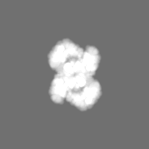
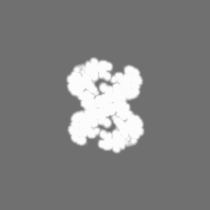
 マップデータ
 試料
試料 機能・相同性情報
 dUTP diphosphatase /
 データ登録者
 英国, 4件
  引用
引用

 構造の表示
構造の表示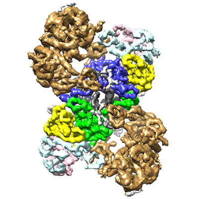
 ダウンロードとリンク
 emd_14860.png
 http://ftp.pdbj.org/pub/emdb/structures/EMD-14860
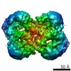


 Z (Sec.)
 Y (Row.)
 X (Col.)


 試料の構成要素
 解析
解析 電子顕微鏡法