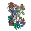
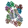
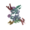
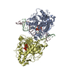
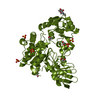
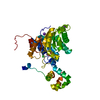
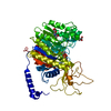
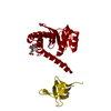
Journal: Science / Year: 2017 Title: A supramolecular assembly mediates lentiviral DNA integration. Authors: Allison Ballandras-Colas / Daniel P Maskell / Erik Serrao / Julia Locke / Paolo Swuec / Stefán R Jónsson / Abhay Kotecha / Nicola J Cook / Valerie E Pye / Ian A Taylor / Valgerdur ...Authors: Allison Ballandras-Colas / Daniel P Maskell / Erik Serrao / Julia Locke / Paolo Swuec / Stefán R Jónsson / Abhay Kotecha / Nicola J Cook / Valerie E Pye / Ian A Taylor / Valgerdur Andrésdóttir / Alan N Engelman / Alessandro Costa / Peter Cherepanov / Abstract: Retroviral integrase (IN) functions within the intasome nucleoprotein complex to catalyze insertion of viral DNA into cellular chromatin. Using cryo-electron microscopy, we now visualize the ...Retroviral integrase (IN) functions within the intasome nucleoprotein complex to catalyze insertion of viral DNA into cellular chromatin. Using cryo-electron microscopy, we now visualize the functional maedi-visna lentivirus intasome at 4.9 angstrom resolution. The intasome comprises a homo-hexadecamer of IN with a tetramer-of-tetramers architecture featuring eight structurally distinct types of IN protomers supporting two catalytically competent subunits. The conserved intasomal core, previously observed in simpler retroviral systems, is formed between two IN tetramers, with a pair of C-terminal domains from flanking tetramers completing the synaptic interface. Our results explain how HIV-1 IN, which self-associates into higher-order multimers, can form a functional intasome, reconcile the bulk of early HIV-1 IN biochemical and structural data, and provide a lentiviral platform for design of HIV-1 IN inhibitors.
History
Deposition
Aug 25, 2016
Deposition site: RCSB / Processing site: PDBE
Revision 1.0
Jan 18, 2017
Provider: repository / Type: Initial release
Revision 2.0
Jan 17, 2024
Group: Atomic model / Data collection ...Atomic model / Data collection / Database references / Refinement description Category: atom_site / chem_comp_atom ...atom_site / chem_comp_atom / chem_comp_bond / database_2 / pdbx_initial_refinement_model Item: _atom_site.occupancy / _database_2.pdbx_DOI / _database_2.pdbx_database_accession
Resolution: 2.501→62.415 Å / SU ML: 0.23 / Cross valid method: FREE R-VALUE / σ(F): 1.36 / Phase error: 26.5 Details: Data were found to be anisotropic, 2.5A in a and c directions; 3.5A in b. Data were subjected to staraniso (global phasing; ian tickle et al) after processing and it is this file the model ...Details: Data were found to be anisotropic, 2.5A in a and c directions; 3.5A in b. Data were subjected to staraniso (global phasing; ian tickle et al) after processing and it is this file the model was refined against (and is uploaded). The model was refined using phenix and coot.
Rfactor
Num. reflection
% reflection
Selection details
Rfree
0.2402
521
5.64 %
random
Rwork
0.1792
-
-
-
obs
0.1823
9236
84.9 %
-
Solvent computation
Shrinkage radii: 0.9 Å / VDW probe radii: 1.11 Å
Displacement parameters
Biso mean: 62.4 Å2
Refinement step
Cycle: LAST / Resolution: 2.501→62.415 Å
Protein
Nucleic acid
Ligand
Solvent
Total
Num. atoms
1716
0
24
46
1786
Refine LS restraints
Refine-ID
Type
Dev ideal
Number
X-RAY DIFFRACTION
f_bond_d
0.002
1792
X-RAY DIFFRACTION
f_angle_d
0.513
2425
X-RAY DIFFRACTION
f_dihedral_angle_d
11.679
1066
X-RAY DIFFRACTION
f_chiral_restr
0.043
258
X-RAY DIFFRACTION
f_plane_restr
0.004
309
LS refinement shell
Resolution (Å)
Rfactor Rfree
Num. reflection Rfree
Rfactor Rwork
Num. reflection Rwork
Refine-ID
% reflection obs (%)
2.5008-2.7525
0.2651
115
0.2111
1493
X-RAY DIFFRACTION
60
2.7525-3.1508
0.2881
120
0.2242
1990
X-RAY DIFFRACTION
79
3.1508-3.9696
0.2477
160
0.1972
2534
X-RAY DIFFRACTION
99
3.9696-62.4344
0.2108
126
0.1542
2698
X-RAY DIFFRACTION
100
Refinement TLS params.
Method: refined / Refine-ID: X-RAY DIFFRACTION
ID
L11 (°2)
L12 (°2)
L13 (°2)
L22 (°2)
L23 (°2)
L33 (°2)
S11 (Å °)
S12 (Å °)
S13 (Å °)
S21 (Å °)
S22 (Å °)
S23 (Å °)
S31 (Å °)
S32 (Å °)
S33 (Å °)
T11 (Å2)
T12 (Å2)
T13 (Å2)
T22 (Å2)
T23 (Å2)
T33 (Å2)
Origin x (Å)
Origin y (Å)
Origin z (Å)
1
0.5452
0.1965
-0.1467
0.5413
0.0338
1.3672
-0.1549
0.0845
-0.121
-0.023
0.0279
0.0097
0.0172
-0.357
-0.0863
0.1677
0.0171
-0.0058
0.2925
-0.014
0.2023
-12.4839
-34.2016
9.3469
2
0.5251
-0.4399
0.1131
1.0577
-0.2091
0.2695
0.0366
0.4469
0.0419
0.0373
-0.1591
-0.1425
0.0546
0.0519
-0.0663
0.1848
-0.0222
0.0172
0.3639
0.0151
0.1666
2.4554
-29.6144
3.025
3
0.0094
-0.0021
-0.0014
0.0039
0.0018
0.0007
0.1854
-0.1921
0.0009
0.1323
0.1624
0.2019
-0.1248
-0.1409
0.0002
0.9106
0.0783
0.0366
0.7224
-0.1528
0.7483
-9.5211
-9.609
21.2039
4
0.0513
-0.0308
0.0683
0.0152
-0.0505
0.1707
0.1609
-0.3983
-0.1937
-0.1882
-0.3811
0.078
-0.1239
0.2146
-0
0.8333
0.0704
0.046
0.8894
-0.013
0.6052
7.6343
-8.1017
38.0689
Refinement TLS group
ID
Refine-ID
Refine TLS-ID
Selection details
1
X-RAY DIFFRACTION
1
(chainAandresid60:153)
2
X-RAY DIFFRACTION
2
(chainAandresid154:207)
3
X-RAY DIFFRACTION
3
(chainAandresid208:220)
4
X-RAY DIFFRACTION
4
(chainAandresid221:276)
+
About Yorodumi
-
News
-
Feb 9, 2022. New format data for meta-information of EMDB entries
New format data for meta-information of EMDB entries
Version 3 of the EMDB header file is now the official format.
The previous official version 1.9 will be removed from the archive.
In the structure databanks used in Yorodumi, some data are registered as the other names, "COVID-19 virus" and "2019-nCoV". Here are the details of the virus and the list of structure data.
Jan 31, 2019. EMDB accession codes are about to change! (news from PDBe EMDB page)
EMDB accession codes are about to change! (news from PDBe EMDB page)
The allocation of 4 digits for EMDB accession codes will soon come to an end. Whilst these codes will remain in use, new EMDB accession codes will include an additional digit and will expand incrementally as the available range of codes is exhausted. The current 4-digit format prefixed with “EMD-” (i.e. EMD-XXXX) will advance to a 5-digit format (i.e. EMD-XXXXX), and so on. It is currently estimated that the 4-digit codes will be depleted around Spring 2019, at which point the 5-digit format will come into force.
The EM Navigator/Yorodumi systems omit the EMD- prefix.
Related info.:Q: What is EMD? / ID/Accession-code notation in Yorodumi/EM Navigator
Yorodumi is a browser for structure data from EMDB, PDB, SASBDB, etc.
This page is also the successor to EM Navigator detail page, and also detail information page/front-end page for Omokage search.
The word "yorodu" (or yorozu) is an old Japanese word meaning "ten thousand". "mi" (miru) is to see.
Related info.:EMDB / PDB / SASBDB / Comparison of 3 databanks / Yorodumi Search / Aug 31, 2016. New EM Navigator & Yorodumi / Yorodumi Papers / Jmol/JSmol / Function and homology information / Changes in new EM Navigator and Yorodumi
 Movie
Movie Controller


 Open data
 Basic information
 Components
 Keywords
 Function and homology information
 Authors
 Citation


 Structure visualization
 Downloads & links
 Other downloads
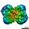


 PDBj
PDBj


 Assembly
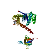


 Mass: 59.044 Da / Num. of mol.: 3 / Source method: obtained synthetically / Formula: C2H3O2
 Mass: 195.237 Da / Num. of mol.: 1 / Source method: obtained synthetically / Formula: C6H13NO4S / Comment: pH buffer*YM
 Mass: 18.015 Da / Num. of mol.: 46 / Source method: isolated from a natural source / Formula: H2O
 Sample preparation
 Processing