Movie
Movie Controller
 Structure viewers
 About Yorodumi Papers


+Search query
-Structure paper
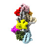
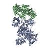
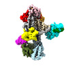
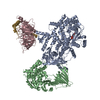
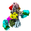
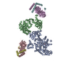
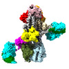
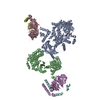
| Title | Molecular basis for Gβγ-mediated activation of phosphoinositide 3-kinase γ. |
|---|---|
| Journal, issue, pages | Nat Struct Mol Biol, Year 2024 |
| Publish date | Apr 2, 2024 |
 Authors Authors | Chun-Liang Chen / Ramizah Syahirah / Sandeep K Ravala / Yu-Chen Yen / Thomas Klose / Qing Deng / John J G Tesmer /  |
| PubMed Abstract | The conversion of phosphatidylinositol 4,5-bisphosphate to phosphatidylinositol 3,4,5-triphosphate by phosphoinositide 3-kinase γ (PI3Kγ) is critical for neutrophil chemotaxis and cancer metastasis. ...The conversion of phosphatidylinositol 4,5-bisphosphate to phosphatidylinositol 3,4,5-triphosphate by phosphoinositide 3-kinase γ (PI3Kγ) is critical for neutrophil chemotaxis and cancer metastasis. PI3Kγ is activated by Gβγ heterodimers released from G protein-coupled receptors responding to extracellular signals. Here we determined cryo-electron microscopy structures of Sus scrofa PI3Kγ-human Gβγ complexes in the presence of substrates/analogs, revealing two Gβγ binding sites: one on the p110γ helical domain and another on the p101 C-terminal domain. Comparison with PI3Kγ alone reveals conformational changes in the kinase domain upon Gβγ binding that are similar to Ras·GTP-induced changes. Assays of variants perturbing the Gβγ binding sites and interdomain contacts altered by Gβγ binding suggest that Gβγ recruits the enzyme to membranes and allosterically regulates activity via both sites. Studies of zebrafish neutrophil migration align with these findings, paving the way for in-depth investigation of Gβγ-mediated activation mechanisms in this enzyme family and drug development for PI3Kγ. |
 External links External links | Nat Struct Mol Biol / PubMed:38565696 |
| Methods | EM (single particle) |
| Resolution | 3.03 - 3.9 Å |
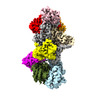
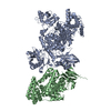
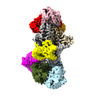
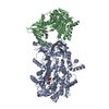
| Structure data | EMDB-40650, PDB-8so9: EMDB-40651, PDB-8soa: EMDB-40652, PDB-8sob: EMDB-40653, PDB-8soc: EMDB-40654, PDB-8sod: EMDB-40655, PDB-8soe: |
| Chemicals |  ChemComp-ATP:  ChemComp-ADP: |
| Source |
|
 Keywords Keywords |  SIGNALING PROTEIN / Phosphoinositide 3-Kinase / Chemotaxis / Cancer SIGNALING PROTEIN / Phosphoinositide 3-Kinase / Chemotaxis / Cancer |