Movie
Movie Controller
 Structure viewers
 About Yorodumi Papers


+Search query
-Structure paper
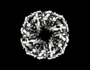
| Title | Design of Diverse Asymmetric Pockets in Homo-oligomeric Proteins. |
|---|---|
| Journal, issue, pages | Biochemistry, Vol. 62, Issue 2, Page 358-368, Year 2023 |
| Publish date | Jan 17, 2023 |
 Authors Authors | Stacey R Gerben / Andrew J Borst / Derrick R Hicks / Isabelle Moczygemba / David Feldman / Brian Coventry / Wei Yang / Asim K Bera / Marcos Miranda / Alex Kang / Hannah Nguyen / David Baker /  |
| PubMed Abstract | A challenge for design of protein-small-molecule recognition is that incorporation of cavities with size, shape, and composition suitable for specific recognition can considerably destabilize protein ...A challenge for design of protein-small-molecule recognition is that incorporation of cavities with size, shape, and composition suitable for specific recognition can considerably destabilize protein monomers. This challenge can be overcome through binding pockets formed at homo-oligomeric interfaces between folded monomers. Interfaces surrounding the central homo-oligomer symmetry axes necessarily have the same symmetry and so may not be well suited to binding asymmetric molecules. To enable general recognition of arbitrary asymmetric substrates and small molecules, we developed an approach to designing asymmetric interfaces at off-axis sites on homo-oligomers, analogous to those found in native homo-oligomeric proteins such as glutamine synthetase. We symmetrically dock curved helical repeat proteins such that they form pockets at the asymmetric interface of the oligomer with sizes ranging from several angstroms, appropriate for binding a single ion, to up to more than 20 Å across. Of the 133 proteins tested, 84 had soluble expression in , 47 had correct oligomeric states in solution, 35 had small-angle X-ray scattering (SAXS) data largely consistent with design models, and 8 had negative-stain electron microscopy (nsEM) 2D class averages showing the structures coming together as designed. Both an X-ray crystal structure and a cryogenic electron microscopy (cryoEM) structure are close to the computational design models. The nature of these proteins as homo-oligomers allows them to be readily built into higher-order structures such as nanocages, and the asymmetric pockets of these structures open rich possibilities for small-molecule binder design free from the constraints associated with monomer destabilization. |
 External links External links | Biochemistry / PubMed:36627259 / PubMed Central |
| Methods | EM (single particle) / X-ray diffraction |
| Resolution | 3.85 - 4.27 Å |
| Structure data | EMDB-27903, PDB-8e55: 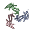 PDB-8e1e: |
| Source |
|
 Keywords Keywords |  DE NOVO PROTEIN / DE NOVO DESIGN / Scaffolding protein / ligand binding / pocket / tetramer / oligomer / pockets / rosetta / cryoEM DE NOVO PROTEIN / DE NOVO DESIGN / Scaffolding protein / ligand binding / pocket / tetramer / oligomer / pockets / rosetta / cryoEM |