 Movie
Movie Controller
 Structure viewers
 About Yorodumi Papers


+Search query
-Structure paper
| Title | In situ architecture of Opa1-dependent mitochondrial cristae remodeling. |
|---|---|
| Journal, issue, pages | EMBO J, Vol. 43, Issue 3, Page 391-413, Year 2024 |
| Publish date | Jan 15, 2024 |
 Authors Authors | Michelle Y Fry / Paula P Navarro / Pusparanee Hakim / Virly Y Ananda / Xingping Qin / Juan C Landoni / Sneha Rath / Zintis Inde / Camila Makhlouta Lugo / Bridget E Luce / Yifan Ge / Julie L McDonald / Ilzat Ali / Leillani L Ha / Benjamin P Kleinstiver / David C Chan / Kristopher A Sarosiek / Luke H Chao /    |
| PubMed Abstract | Cristae membrane state plays a central role in regulating mitochondrial function and cellular metabolism. The protein Optic atrophy 1 (Opa1) is an important crista remodeler that exists as two forms ...Cristae membrane state plays a central role in regulating mitochondrial function and cellular metabolism. The protein Optic atrophy 1 (Opa1) is an important crista remodeler that exists as two forms in the mitochondrion, a membrane-anchored long form (l-Opa1) and a processed short form (s-Opa1). The mechanisms for how Opa1 influences cristae shape have remained unclear due to lack of native three-dimensional views of cristae. We perform in situ cryo-electron tomography of cryo-focused ion beam milled mouse embryonic fibroblasts with defined Opa1 states to understand how each form of Opa1 influences cristae architecture. In our tomograms, we observe a variety of cristae shapes with distinct trends dependent on s-Opa1:l-Opa1 balance. Increased l-Opa1 levels promote cristae stacking and elongated mitochondria, while increased s-Opa1 levels correlated with irregular cristae packing and round mitochondria shape. Functional assays indicate a role for l-Opa1 in wild-type apoptotic and calcium handling responses, and show a compromised respiratory function under Opa1 imbalance. In summary, we provide three-dimensional visualization of cristae architecture to reveal relationships between mitochondrial ultrastructure and cellular function dependent on Opa1-mediated membrane remodeling. |
 External links External links | EMBO J / PubMed:38225406 / PubMed Central |
| Methods | EM (tomography) |
| Structure data | 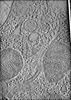 EMDB-43049: Cryo-electron tomogram of mitochondria from cryo-FIB milled l-Opa1* mouse embryonic fibroblasts 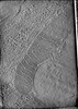 EMDB-43050: cryo-electron tomogram of mitochondria in cryo-FIB milled l-Opa1* mouse embryonic fibroblasts  EMDB-43051: cryo-electron tomogram of mitochondria in cryo-FIB milled l-Opa1* mouse embryonic fibroblasts 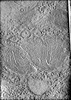 EMDB-43052: cryo-electron tomogram of mitochondria in cryo-FIB milled l-Opa1* mouse embryonic fibroblasts  EMDB-43053: Cryo-electron tomogram of mitochondria in cryo-FIB milled s-Opa1* mouse embryonic fibroblasts  EMDB-43054: cryo-electron tomogram of mitochondria in cryo-FIB milled s-Opa1* mouse embryonic fibroblasts  EMDB-43055: cryo-electron tomogram of mitochondria in cryo-FIB milled s-Opa1* mouse embryonic fibroblasts  EMDB-43056: cryo-electron tomogram of mitochondria in cryo-FIB milled s-Opa1* mouse embryonic fibroblasts 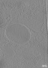 EMDB-43057: Cryo-electron tomogram of mitochondria in cryo-FIB milled s-Opa1* mouse embryonic fibroblasts  EMDB-43058: Cryo-electron tomogram of mitochondria in cryo-FIB milled mouse embryonic fibroblasts with OPA1 deletion  EMDB-43059: Cryo-electron tomogram of mitochondria in cryo-FIB milled mouse embryonic fibroblasts with OPA1 deletion  EMDB-43060: Cryo-electron tomogram of mitochondria in cryo-FIB milled mouse embryonic fibroblasts with OPA1 deletion  EMDB-43061: Cryo-electron tomogram of mitochondria in cryo-FIB milled mouse embryonic fibroblasts with OPA1 deletion  EMDB-43062: Cryo-electron tomogram of mitochondria in cryo-FIB milled mouse embryonic fibroblasts with OPA1 deletion  EMDB-43063: Cryo-electron tomogram of mitochondria in cryo-FIB milled mouse embryonic fibroblasts  EMDB-43064: Cryo-electron tomogram of mitochondria in cryo-FIB milled mouse embryonic fibroblasts  EMDB-43065: Cryo-electron tomogram of mitochondria in cryo-FIB milled mouse embryonic fibroblasts  EMDB-43066: Cryo-electron tomogram of mitochondria in cryo-FIB milled mouse embryonic fibroblasts  EMDB-43067: Cryo-electron tomogram of mitochondria in cryo-FIB milled mouse embryonic fibroblasts with overexpression of OPA1  EMDB-43068: Cryo-electron tomogram of mitochondria in cryo-FIB milled mouse embryonic fibroblasts with overexpression of OPA1  EMDB-43069: Cryo-electron tomogram of mitochondria in cryo-FIB milled mouse embryonic fibroblasts with overexpression of OPA1  EMDB-43070: Cryo-electron tomogram of mitochondria in cryo-FIB milled mouse embryonic fibroblasts with overexpression of OPA1 |
| Source |
|

 Mus musculus (house mouse)