National Institutes of Health/National Institute of General Medical Sciences (NIH/NIGMS)
GM142553
United States
National Institutes of Health/National Institute of Diabetes and Digestive and Kidney Disease (NIH/NIDDK)
DK125263
United States
National Institutes of Health/National Cancer Institute (NIH/NCI)
CA248565
United States
Jane Coffin Childs (JCC) Fund
United States
Swiss National Science Foundation
P2BSP3_188112
Switzerland
Swiss National Science Foundation
P400PB_199252
Switzerland
Citation
Journal: EMBO J / Year: 2024 Title: In situ architecture of Opa1-dependent mitochondrial cristae remodeling. Authors: Michelle Y Fry / Paula P Navarro / Pusparanee Hakim / Virly Y Ananda / Xingping Qin / Juan C Landoni / Sneha Rath / Zintis Inde / Camila Makhlouta Lugo / Bridget E Luce / Yifan Ge / Julie L ...Authors: Michelle Y Fry / Paula P Navarro / Pusparanee Hakim / Virly Y Ananda / Xingping Qin / Juan C Landoni / Sneha Rath / Zintis Inde / Camila Makhlouta Lugo / Bridget E Luce / Yifan Ge / Julie L McDonald / Ilzat Ali / Leillani L Ha / Benjamin P Kleinstiver / David C Chan / Kristopher A Sarosiek / Luke H Chao / Abstract: Cristae membrane state plays a central role in regulating mitochondrial function and cellular metabolism. The protein Optic atrophy 1 (Opa1) is an important crista remodeler that exists as two forms ...Cristae membrane state plays a central role in regulating mitochondrial function and cellular metabolism. The protein Optic atrophy 1 (Opa1) is an important crista remodeler that exists as two forms in the mitochondrion, a membrane-anchored long form (l-Opa1) and a processed short form (s-Opa1). The mechanisms for how Opa1 influences cristae shape have remained unclear due to lack of native three-dimensional views of cristae. We perform in situ cryo-electron tomography of cryo-focused ion beam milled mouse embryonic fibroblasts with defined Opa1 states to understand how each form of Opa1 influences cristae architecture. In our tomograms, we observe a variety of cristae shapes with distinct trends dependent on s-Opa1:l-Opa1 balance. Increased l-Opa1 levels promote cristae stacking and elongated mitochondria, while increased s-Opa1 levels correlated with irregular cristae packing and round mitochondria shape. Functional assays indicate a role for l-Opa1 in wild-type apoptotic and calcium handling responses, and show a compromised respiratory function under Opa1 imbalance. In summary, we provide three-dimensional visualization of cristae architecture to reveal relationships between mitochondrial ultrastructure and cellular function dependent on Opa1-mediated membrane remodeling.
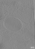
Download / File: emd_43049.map.gz / Format: CCP4 / Size: 315.8 MB / Type: IMAGE STORED AS FLOATING POINT NUMBER (4 BYTES)
Voxel size
X=Y=Z: 22.06 Å
Density
Minimum - Maximum
-32.206435999999997 - 87.248069999999998
Average (Standard dev.)
13.352292 (±2.9149015)
Symmetry
Space group: 1
Details
EMDB XML:
Map geometry
Axis order
X
Y
Z
Origin
-104
104
-113
Dimensions
720
511
225
Spacing
511
720
225
Cell
A: 11272.66 Å / B: 15883.199 Å / C: 4963.5 Å α=β=γ: 90.0 °
-
Supplemental data
-
Sample components
-
Entire : mitochondria in mouse embryonic fibroblasts with OMA1 deletion an...
Entire
Name: mitochondria in mouse embryonic fibroblasts with OMA1 deletion and OPA1-exon5b deletionMitochondrion
Components
Cell: mitochondria in mouse embryonic fibroblasts with OMA1 deletion and OPA1-exon5b deletionMitochondrion
-
Supramolecule #1: mitochondria in mouse embryonic fibroblasts with OMA1 deletion an...
Supramolecule
Name: mitochondria in mouse embryonic fibroblasts with OMA1 deletion and OPA1-exon5b deletion type: cell / ID: 1 / Parent: 0
Source (natural)
Organism: Mus musculus (house mouse)
-
Experimental details
-
Structure determination
Method
cryo EM
Processing
electron tomography
Aggregation state
cell
-
Sample preparation
Buffer
pH: 7.5
Vitrification
Cryogen name: ETHANE / Chamber humidity: 100 % / Chamber temperature: 277.15 K / Instrument: FEI VITROBOT MARK IV
Sectioning
Focused ion beam - Instrument: OTHER / Focused ion beam - Ion: OTHER / Focused ion beam - Voltage: 5 / Focused ion beam - Current: 0.01 / Focused ion beam - Duration: 600 / Focused ion beam - Temperature: 88 K / Focused ion beam - Initial thickness: 1000 / Focused ion beam - Final thickness: 150 Focused ion beam - Details: The value given for _em_focused_ion_beam.instrument is TFS Aquilos2. This is not in a list of allowed values {'DB235', 'OTHER'} so OTHER is written into the XML file.
-
Electron microscopy
Microscope
FEI TITAN KRIOS
Electron beam
Acceleration voltage: 300 kV / Electron source: FIELD EMISSION GUN
In the structure databanks used in Yorodumi, some data are registered as the other names, "COVID-19 virus" and "2019-nCoV". Here are the details of the virus and the list of structure data.
Jan 31, 2019. EMDB accession codes are about to change! (news from PDBe EMDB page)
EMDB accession codes are about to change! (news from PDBe EMDB page)
The allocation of 4 digits for EMDB accession codes will soon come to an end. Whilst these codes will remain in use, new EMDB accession codes will include an additional digit and will expand incrementally as the available range of codes is exhausted. The current 4-digit format prefixed with “EMD-” (i.e. EMD-XXXX) will advance to a 5-digit format (i.e. EMD-XXXXX), and so on. It is currently estimated that the 4-digit codes will be depleted around Spring 2019, at which point the 5-digit format will come into force.
The EM Navigator/Yorodumi systems omit the EMD- prefix.
Related info.:Q: What is EMD? / ID/Accession-code notation in Yorodumi/EM Navigator
Yorodumi is a browser for structure data from EMDB, PDB, SASBDB, etc.
This page is also the successor to EM Navigator detail page, and also detail information page/front-end page for Omokage search.
The word "yorodu" (or yorozu) is an old Japanese word meaning "ten thousand". "mi" (miru) is to see.
Related info.:EMDB / PDB / SASBDB / Comparison of 3 databanks / Yorodumi Search / Aug 31, 2016. New EM Navigator & Yorodumi / Yorodumi Papers / Jmol/JSmol / Function and homology information / Changes in new EM Navigator and Yorodumi
 Movie
Movie Controller

 Yorodumi
 Open data
 Basic information
 Map data
 Sample
 Keywords
 mitochondria / cristae /
 Authors
 United States,
  Switzerland, 6 items
  Citation
 Structure visualization
 Downloads & links
 EMDB map data format
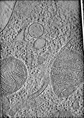 emd_43049.png
 http://ftp.pdbj.org/pub/emdb/structures/EMD-43049


 Sample components
 Processing
 Electron microscopy
