Movie
Movie Controller
 Structure viewers
 About Yorodumi Papers


+Search query
-Structure paper
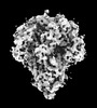
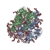
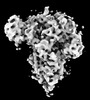
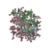
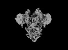
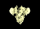
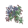
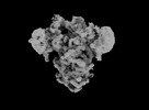
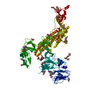
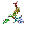
| Title | In situ structure and dynamics of an alphacoronavirus spike protein by cryo-ET and cryo-EM. |
|---|---|
| Journal, issue, pages | Nat Commun, Vol. 13, Issue 1, Page 4877, Year 2022 |
| Publish date | Aug 19, 2022 |
 Authors Authors | Cheng-Yu Huang / Piotr Draczkowski / Yong-Sheng Wang / Chia-Yu Chang / Yu-Chun Chien / Yun-Han Cheng / Yi-Min Wu / Chun-Hsiung Wang / Yuan-Chih Chang / Yen-Chen Chang / Tzu-Jing Yang / Yu-Xi Tsai / Kay-Hooi Khoo / Hui-Wen Chang / Shang-Te Danny Hsu /   |
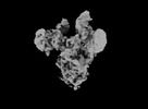
| PubMed Abstract | Porcine epidemic diarrhea (PED) is a highly contagious swine disease caused by porcine epidemic diarrhea virus (PEDV). PED causes enteric disorders with an exceptionally high fatality in neonates, ...Porcine epidemic diarrhea (PED) is a highly contagious swine disease caused by porcine epidemic diarrhea virus (PEDV). PED causes enteric disorders with an exceptionally high fatality in neonates, bringing substantial economic losses in the pork industry. The trimeric spike (S) glycoprotein of PEDV is responsible for virus-host recognition, membrane fusion, and is the main target for vaccine development and antigenic analysis. The atomic structures of the recombinant PEDV S proteins of two different strains have been reported, but they reveal distinct N-terminal domain 0 (D0) architectures that may correspond to different functional states. The existence of the D0 is a unique feature of alphacoronavirus. Here we combined cryo-electron tomography (cryo-ET) and cryo-electron microscopy (cryo-EM) to demonstrate in situ the asynchronous S protein D0 motions on intact viral particles of a highly virulent PEDV Pintung 52 strain. We further determined the cryo-EM structure of the recombinant S protein derived from a porcine cell line, which revealed additional domain motions likely associated with receptor binding. By integrating mass spectrometry and cryo-EM, we delineated the complex compositions and spatial distribution of the PEDV S protein N-glycans, and demonstrated the functional role of a key N-glycan in modulating the D0 conformation. |
 External links External links | Nat Commun / PubMed:35986008 / PubMed Central |
| Methods | EM (single particle) / EM (subtomogram averaging) |
| Resolution | 3.1 - 31.0 Å |
| Structure data | EMDB-32329, PDB-7w6m: 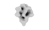 EMDB-32332: Subtomogram averaging of PEDV (Pintung 52) S protein with all three protomers in the D0-down conformation determined in situ on intact viral particles. 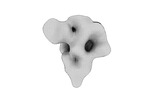 EMDB-32333: Subtomogram averaging of PEDV (Pintung 52) S protein with one protomer in the D0-up conformation and two protomers in the D0-down conformation, determined in situ on intact viral particles 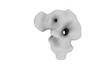 EMDB-32337: Subtomogram averaging of PEDV (Pintung 52) S protein with two protomers in the D0-up conformation and one protomer in the D0-down conformation, determined in situ on intact viral particles. EMDB-32338, PDB-7w73: 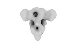 EMDB-32339: Subtomogram averaging of PEDV (Pintung 52) S protein with all three protomers in the D0-up conformation determined in situ on intact viral particles. 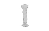 EMDB-32340: Subtomogram averaging of PEDV (Pintung 52) S protein in the postfusion form determined in situ on intact viral particles. EMDB-33646, PDB-7y6s: EMDB-33647, PDB-7y6t: EMDB-33648, PDB-7y6u: EMDB-33649, PDB-7y6v: 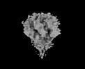 EMDB-33700: Cryo-EM map of HEK293F cell-derived PEDV PT52 S protein with three D0-down 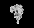 EMDB-33701: Cryo-EM map of HEK293F cell-derived PEDV PT52 S protein one D0-up and two D0-down 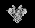 EMDB-33702: Cryo-EM map of HEK293F cell-derived PEDV PT52 S protein with three D0-up 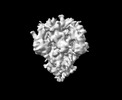 EMDB-33703: Cryo-EM map of HEK293F cell-derived PEDV PT52 S T326I with three D0-down 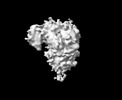 EMDB-33704: Cryo-EM map of HEK293F cell-derived PEDV PT52 S T326I one D0-up and two D0-down 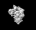 EMDB-33705: Cryo-EM map of HEK293F cell-derived PEDV PT52 S T326I one D0-down and two D0-up 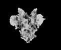 EMDB-33706: Cryo-EM map of HEK293F cell-derived PEDV PT52 S T326I with three D0-up |
| Chemicals |  ChemComp-NAG: |
| Source |
|
 Keywords Keywords |  VIRAL PROTEIN / PEDV / Spike / Glycoprotein VIRAL PROTEIN / PEDV / Spike / Glycoprotein |