 Movie
Movie Controller

[English] 日本語
 Yorodumi
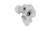
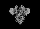
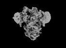
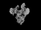
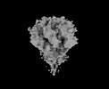
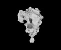
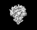
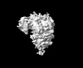
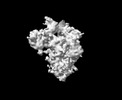
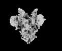
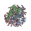
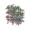
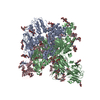
- EMDB-33702: Cryo-EM map of HEK293F cell-derived PEDV PT52 S protein with thre... -
+ Open data

- Basic information
| Entry |  | |||||||||||||||||||||
|---|---|---|---|---|---|---|---|---|---|---|---|---|---|---|---|---|---|---|---|---|---|---|
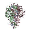
| Title | Cryo-EM map of HEK293F cell-derived PEDV PT52 S protein with three D0-up | |||||||||||||||||||||
 Map data Map data | ||||||||||||||||||||||
 Sample Sample |
| |||||||||||||||||||||
| Biological species |   Porcine epidemic diarrhea virus Porcine epidemic diarrhea virus | |||||||||||||||||||||
| Method | single particle reconstruction / cryo EM / Resolution: 4.5 Å | |||||||||||||||||||||
 Authors Authors | Hsu STD / Draczkowski P / Wang YS | |||||||||||||||||||||
| Funding support |  Taiwan, 6 items Taiwan, 6 items
| |||||||||||||||||||||
 Citation Citation | Journal: Nat Commun / Year: 2022 Title: In situ structure and dynamics of an alphacoronavirus spike protein by cryo-ET and cryo-EM. Authors: Cheng-Yu Huang / Piotr Draczkowski / Yong-Sheng Wang / Chia-Yu Chang / Yu-Chun Chien / Yun-Han Cheng / Yi-Min Wu / Chun-Hsiung Wang / Yuan-Chih Chang / Yen-Chen Chang / Tzu-Jing Yang / Yu-Xi ...Authors: Cheng-Yu Huang / Piotr Draczkowski / Yong-Sheng Wang / Chia-Yu Chang / Yu-Chun Chien / Yun-Han Cheng / Yi-Min Wu / Chun-Hsiung Wang / Yuan-Chih Chang / Yen-Chen Chang / Tzu-Jing Yang / Yu-Xi Tsai / Kay-Hooi Khoo / Hui-Wen Chang / Shang-Te Danny Hsu /  Abstract: Porcine epidemic diarrhea (PED) is a highly contagious swine disease caused by porcine epidemic diarrhea virus (PEDV). PED causes enteric disorders with an exceptionally high fatality in neonates, ...Porcine epidemic diarrhea (PED) is a highly contagious swine disease caused by porcine epidemic diarrhea virus (PEDV). PED causes enteric disorders with an exceptionally high fatality in neonates, bringing substantial economic losses in the pork industry. The trimeric spike (S) glycoprotein of PEDV is responsible for virus-host recognition, membrane fusion, and is the main target for vaccine development and antigenic analysis. The atomic structures of the recombinant PEDV S proteins of two different strains have been reported, but they reveal distinct N-terminal domain 0 (D0) architectures that may correspond to different functional states. The existence of the D0 is a unique feature of alphacoronavirus. Here we combined cryo-electron tomography (cryo-ET) and cryo-electron microscopy (cryo-EM) to demonstrate in situ the asynchronous S protein D0 motions on intact viral particles of a highly virulent PEDV Pintung 52 strain. We further determined the cryo-EM structure of the recombinant S protein derived from a porcine cell line, which revealed additional domain motions likely associated with receptor binding. By integrating mass spectrometry and cryo-EM, we delineated the complex compositions and spatial distribution of the PEDV S protein N-glycans, and demonstrated the functional role of a key N-glycan in modulating the D0 conformation. | |||||||||||||||||||||
| History |
|
- Structure visualization
| Supplemental images |
|---|

- Downloads & links
-EMDB archive
| Map data | emd_33702.map.gz | 11.6 MB |  EMDB map data format EMDB map data format | |
|---|---|---|---|---|
| Header (meta data) | emd-33702-v30.xmlemd-33702.xml | 20.1 KB 20.1 KB | Display Display | EMDB header |
| FSC (resolution estimation) | emd_33702_fsc.xml | 6 KB | Display | FSC data file |
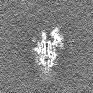
| Images | 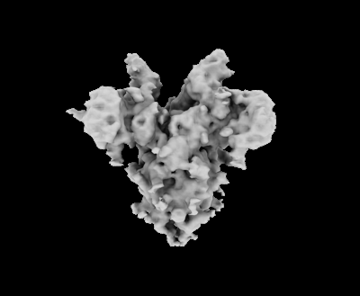 emd_33702.png emd_33702.png | 35.8 KB | ||
| Others | emd_33702_half_map_1.map.gzemd_33702_half_map_2.map.gz | 21.4 MB 21.4 MB | ||
| Archive directory |  http://ftp.pdbj.org/pub/emdb/structures/EMD-33702ftp://ftp.pdbj.org/pub/emdb/structures/EMD-33702 http://ftp.pdbj.org/pub/emdb/structures/EMD-33702ftp://ftp.pdbj.org/pub/emdb/structures/EMD-33702 | HTTPS FTP |
-Related structure data

-Links
| EMDB pages | EMDB (EBI/PDBe) / EMDataResource |
|---|
-Map
| File | Download / File: emd_33702.map.gz / Format: CCP4 / Size: 23 MB / Type: IMAGE STORED AS FLOATING POINT NUMBER (4 BYTES) | ||||||||||||||||||||
|---|---|---|---|---|---|---|---|---|---|---|---|---|---|---|---|---|---|---|---|---|---|
| Voxel size | X=Y=Z: 2.2 Å | ||||||||||||||||||||
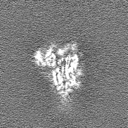
| Density |
| ||||||||||||||||||||
| Symmetry | Space group: 1 | ||||||||||||||||||||
| Details | EMDB XML:
|
-Supplemental data
-Half map: #1
| File | emd_33702_half_map_1.map | ||||||||||||
|---|---|---|---|---|---|---|---|---|---|---|---|---|---|
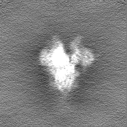
| Projections & Slices |
| ||||||||||||
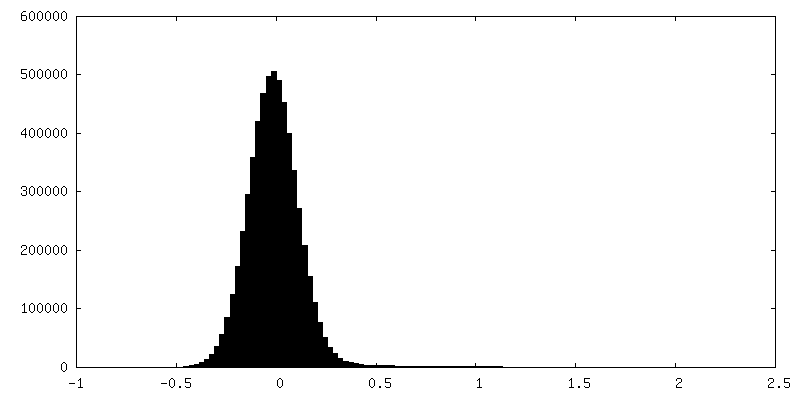
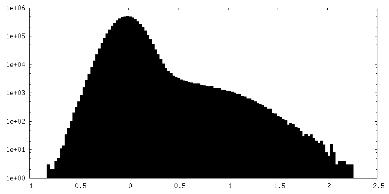
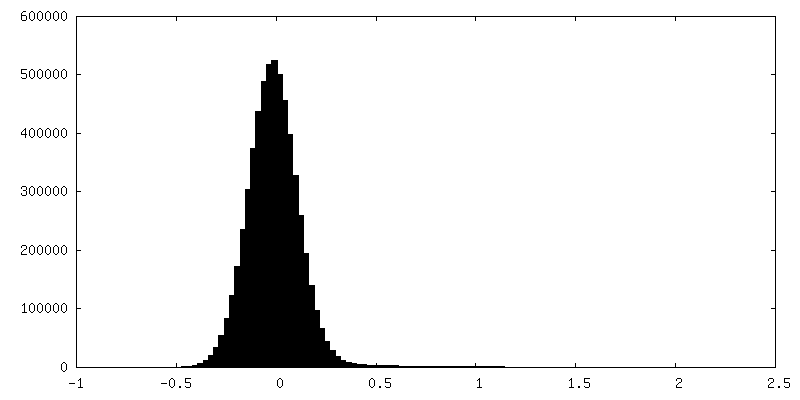
| Density Histograms |
 Z
Z Y
Y X
X


-Half map: #2
| File | emd_33702_half_map_2.map | ||||||||||||
|---|---|---|---|---|---|---|---|---|---|---|---|---|---|
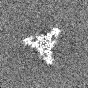
| Projections & Slices |
| ||||||||||||
| Density Histograms |


- Sample components
-Entire : recombinant PEDV PT52 S protein expressed from HEK293F cells
| Entire | Name: recombinant PEDV PT52 S protein expressed from HEK293F cells |
|---|---|
| Components |
|
-Supramolecule #1: recombinant PEDV PT52 S protein expressed from HEK293F cells
| Supramolecule | Name: recombinant PEDV PT52 S protein expressed from HEK293F cells type: organelle_or_cellular_component / ID: 1 / Parent: 0 / Macromolecule list: all |
|---|---|
| Source (natural) | Organism: Porcine epidemic diarrhea virus / Strain: Pintung 52 |
| Recombinant expression | Organism:  Homo sapiens (human) / Recombinant cell: HEK293F Homo sapiens (human) / Recombinant cell: HEK293F |
-Macromolecule #1: recombinant PEDV PT52 S protein expressed from HEK293F cells
| Macromolecule | Name: recombinant PEDV PT52 S protein expressed from HEK293F cells type: protein_or_peptide / ID: 1 / Enantiomer: LEVO |
|---|---|
| Source (natural) | Organism: Porcine epidemic diarrhea virus / Strain: Pintung52 |
| Recombinant expression | Organism: Homo sapiens (human) |
| Sequence | String: MKSLTYFWLF LPVLSTLSLP QDVTRCSAKT NFRRFFSKFN VQAPAVVVLG GYLPIGENQG VNSTWYCAGQ HPTASGVHGI FVSHIRGGHG FEIGISQEPF DPSGYQLYLH KATNGNTNAT ARLRICQFPS IKTLGPTANN DVTIGRNCLF NKAIPAHMSE HSVVGITWDN ...String: MKSLTYFWLF LPVLSTLSLP QDVTRCSAKT NFRRFFSKFN VQAPAVVVLG GYLPIGENQG VNSTWYCAGQ HPTASGVHGI FVSHIRGGHG FEIGISQEPF DPSGYQLYLH KATNGNTNAT ARLRICQFPS IKTLGPTANN DVTIGRNCLF NKAIPAHMSE HSVVGITWDN DRVTVFSDKI YYFYFKNDWS RVATKCYNSG GCAMQYVYEP TYYMLNVTSA GEDGISYQPC TANCIGYAAN VFATEPNGHI PEGFSFNNWF LLSNDSTLVH GKVVSNQPLL VNCLLAIPKI YGLGQFFSFN QTIDGVCNGA AVQRAPEALR FNINDTSVIL AEGSIVLHTA LGTNFSFVCS NSSNPHLATF AIPLGATQVP YYCFFKVDTY NSTVYKFLAV LPPTVREIVI TKYGDVYVNG FGYLHLGLLD AVTINFTGHG TDDDVSGFWT IASTNFVDAL IEVQGTAIQR ILYCDDPVSQ LKCSQVAFDL DDGFYPFSSR NLLSHEQPIS FVTLPSFNAH SFVNITVSAS FGGHSGANLI ASDTTINGFS SFCVDTRQFT ISLSYNVTNS YGYVSNSQDS NCPFTLQSVN DYLSFSKFCV STSLLASACT IDLFGYPEFG SGVKFTSLYF QFTKGELITG TPKPLEGVTD VSFMTLDVCT KYTIYGFKGE GIITLTNSSF LAGVYYTSDS GQLLAFKNVT SGAVYSVTPC SFSEQAAYVD DDIVGVISSL SSSTFNSTRE LPGFFYHSND GSNCTEPVLV YSNIGVCKSG SIGYVPSQSG QVKIAPTVTG NISIPTNFSM SIRTEYLQLY NTPVSVDCAT YVCNGNSRCK QLLTQYTAAC KTIESALQLS ARLESVEVNS MLTISEEALQ LATISSFNGD GYNFTNVLGV SVYDPARGRV VQKRSFIEDL LFNKVVTNGL GTVDEDYKRC SNGRSVADLV CAQYYSGVMV LPGVVDAEKL HMYSASLIGG MVLGGFTAAA ALPFSYAVQA RLNYLALQTD VLQRNQQLLA ESFNSAIGNI TSAFESVKEA SSQTSRGLNT VAHALTKVQE VVNSQGAALT QLTVQLQHNF QAISSSIDDI YSRLDPPSAD VQVDRLITGR LSALNAFVAQ TLTKYTEVQA SRKLAQQKVN ECVKSQSQRY GFCGGDGEHI FSLVQAAPQG LLFLHTVLVP SDFVDVIAIA GLCVNDEIAL TLREPGLVLF THELQNHTAT EYFVSSRRMF EPRKPTVSDF VQIESCVVTY VNLTRDQLPD VIPDYIDVNK TRDEILASLP NRTGPSLPLD VFNATYLNLT GEIADLEQRS ESLRNTTEEL QSLIYNINNT LVDLEWLNRV ETYIKWPEFG SGGYIPEAPR DGQAYVRKDG EWVLLSTFLK GQDNSADIQH SGRPLESRGP FEQKLISEED LNMHTGHHHH HH |
-Experimental details
-Structure determination
| Method | cryo EM |
|---|---|
 Processing Processing | single particle reconstruction |
| Aggregation state | particle |
-Sample preparation
| Buffer | pH: 7.6 Component:
Details: Blot for 3 seconds before plunging. Force 0. | ||||||||||||
|---|---|---|---|---|---|---|---|---|---|---|---|---|---|
| Vitrification | Cryogen name: ETHANE / Chamber humidity: 100 % / Chamber temperature: 277.15 K / Instrument: FEI VITROBOT MARK IV | ||||||||||||
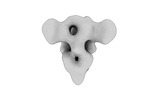
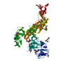
| Details | Cryo-EM map of HEK293F cell-derived PEDV PT52 S protein with three D0-down |
- Electron microscopy
| Microscope | FEI TITAN KRIOS |
|---|---|
| Electron beam | Acceleration voltage: 300 kV / Electron source: FIELD EMISSION GUN |
| Electron optics | C2 aperture diameter: 100.0 µm / Illumination mode: FLOOD BEAM / Imaging mode: BRIGHT FIELDBright-field microscopy / Cs: 2.7 mm / Nominal defocus max: 2.3000000000000003 µm / Nominal defocus min: 1.8 µm / Nominal magnification: 81000 |
| Specialist optics | Energy filter - Name: GIF Bioquantum |
| Sample stage | Specimen holder model: FEI TITAN KRIOS AUTOGRID HOLDER / Cooling holder cryogen: NITROGEN |
| Details | Data were recorded with stage tilt at 0 and 30 degree |
| Image recording | Film or detector model: GATAN K3 (6k x 4k) / Number grids imaged: 1 / Number real images: 2574 / Average electron dose: 50.0 e/Å2 |
| Experimental equipment |  Model: Titan Krios / Image courtesy: FEI Company |
-Image processing
| Particle selection | Number selected: 337583 |
|---|---|
| CTF correction | Software - Name: CTFFIND (ver. 4.1) |
| Startup model | Type of model: PDB ENTRY PDB model - PDB ID: |
| Initial angle assignment | Type: MAXIMUM LIKELIHOOD / Software - Name: RELION (ver. 3.1) |
| Final 3D classification | Details: Non-uniform refinement in CryoSparc3.1 |
| Final angle assignment | Type: OTHER / Software - Name: cryoSPARC (ver. 3.1) / Details: Non-uniform refinement in CryoSparc3.1 |
| Final reconstruction | Resolution.type: BY AUTHOR / Resolution: 4.5 Å / Resolution method: FSC 0.143 CUT-OFF / Software - Name: cryoSPARC (ver. 3.1) / Number images used: 78249 |
| FSC plot (resolution estimation) |  |
-Atomic model buiding 1
| Refinement | Space: REAL / Protocol: RIGID BODY FIT |
|---|
