 Movie
Movie Controller
 Structure viewers
 About Yorodumi Papers


+Search query
-Structure paper
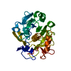
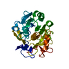
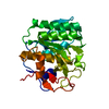
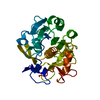
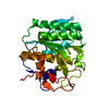
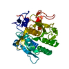
| Title | Benchmarking the ideal sample thickness in cryo-EM. |
|---|---|
| Journal, issue, pages | Proc Natl Acad Sci U S A, Vol. 118, Issue 49, Year 2021 |
| Publish date | Dec 7, 2021 |
 Authors Authors | Michael W Martynowycz / Max T B Clabbers / Johan Unge / Johan Hattne / Tamir Gonen /  |
| PubMed Abstract | The relationship between sample thickness and quality of data obtained is investigated by microcrystal electron diffraction (MicroED). Several electron microscopy (EM) grids containing proteinase K ...The relationship between sample thickness and quality of data obtained is investigated by microcrystal electron diffraction (MicroED). Several electron microscopy (EM) grids containing proteinase K microcrystals of similar sizes from the same crystallization batch were prepared. Each grid was transferred into a focused ion beam and a scanning electron microscope in which the crystals were then systematically thinned into lamellae between 95- and 1,650-nm thick. MicroED data were collected at either 120-, 200-, or 300-kV accelerating voltages. Lamellae thicknesses were expressed in multiples of the corresponding inelastic mean free path to allow the results from different acceleration voltages to be compared. The quality of the data and subsequently determined structures were assessed using standard crystallographic measures. Structures were reliably determined with similar quality from crystalline lamellae up to twice the inelastic mean free path. Lower resolution diffraction was observed at three times the mean free path for all three accelerating voltages, but the data quality was insufficient to yield structures. Finally, no coherent diffraction was observed from lamellae thicker than four times the calculated inelastic mean free path. This study benchmarks the ideal specimen thickness with implications for all cryo-EM methods. |
 External links External links | Proc Natl Acad Sci U S A / PubMed:34873060 / PubMed Central |
| Methods | EM (electron crystallography) |
| Resolution | 1.85 - 2.9 Å |
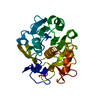
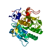
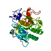
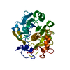
| Structure data | EMDB-25456, PDB-7svy: EMDB-25457, PDB-7svz: EMDB-25458, PDB-7sw0: EMDB-25459, PDB-7sw1: EMDB-25460, PDB-7sw2: EMDB-25461, PDB-7sw3: EMDB-25462, PDB-7sw4: EMDB-25463, PDB-7sw5: EMDB-25464, PDB-7sw6: EMDB-25465, PDB-7sw7: EMDB-25466, PDB-7sw8: EMDB-25467, PDB-7sw9: EMDB-25468, PDB-7swa: EMDB-25469, PDB-7swb: EMDB-25470, PDB-7swc: |
| Chemicals |  ChemComp-HOH: |
| Source |
|
 Keywords Keywords |  HYDROLASE / Serine protease HYDROLASE / Serine protease |


 parengyodontium album (fungus)