Movie
Movie Controller
 Structure viewers
 About Yorodumi Papers


+Search query
-Structure paper
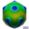
| Title | The giant mimivirus 1.2 Mb genome is elegantly organized into a 30-nm diameter helical protein shield. |
|---|---|
| Journal, issue, pages | Elife, Vol. 11, Year 2022 |
| Publish date | Jul 28, 2022 |
 Authors Authors | Alejandro Villalta / Alain Schmitt / Leandro F Estrozi / Emmanuelle R J Quemin / Jean-Marie Alempic / Audrey Lartigue / Vojtěch Pražák / Lucid Belmudes / Daven Vasishtan / Agathe M G Colmant / Flora A Honoré / Yohann Couté / Kay Grünewald / Chantal Abergel /    |
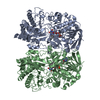
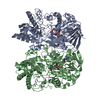
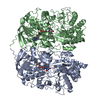
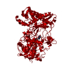
| PubMed Abstract | Mimivirus is the prototype of the family of giant dsDNA viruses. Little is known about the organization of the 1.2 Mb genome inside the membrane-limited nucleoid filling the ~0.5 µm icosahedral ...Mimivirus is the prototype of the family of giant dsDNA viruses. Little is known about the organization of the 1.2 Mb genome inside the membrane-limited nucleoid filling the ~0.5 µm icosahedral capsids. Cryo-electron microscopy, cryo-electron tomography, and proteomics revealed that it is encased into a ~30-nm diameter helical protein shell surprisingly composed of two GMC-type oxidoreductases, which also form the glycosylated fibrils decorating the capsid. The genome is arranged in 5- or 6-start left-handed super-helices, with each DNA-strand lining the central channel. This luminal channel of the nucleoprotein fiber is wide enough to accommodate oxidative stress proteins and RNA polymerase subunits identified by proteomics. Such elegant supramolecular organization would represent a remarkable evolutionary strategy for packaging and protecting the genome, in a state ready for immediate transcription upon unwinding in the host cytoplasm. The parsimonious use of the same protein in two unrelated substructures of the virion is unexpected for a giant virus with thousand genes at its disposal. |
 External links External links | Elife / PubMed:35900198 / PubMed Central |
| Methods | EM (single particle) / EM (helical sym.) / EM (tomography) |
| Resolution | 3.3 - 4.0 Å |
| Structure data | EMDB-13641, PDB-7ptv: EMDB-14353, PDB-7yx3: EMDB-14354, PDB-7yx4: EMDB-14355, PDB-7yx5:  EMDB-15627: Tomogram of the Mimivirus genomic fiber  EMDB-15628: Tomogram of the Mimivirus genomic fiber  EMDB-15629: Tomogram of the Mimivirus genomic fiber  EMDB-15630: Tomogram of the Mimivirus genomic fiber |
| Chemicals |  ChemComp-FAD: |
| Source |
|
 Keywords Keywords |  VIRAL PROTEIN / Mimivirus / Genomic fibre / Cytoplasmic infectious cycle / 1.2 Mb dsDNA / VIRUS VIRAL PROTEIN / Mimivirus / Genomic fibre / Cytoplasmic infectious cycle / 1.2 Mb dsDNA / VIRUS |