Movie
Movie Controller
 Structure viewers
 About Yorodumi Papers


+Search query
-Structure paper
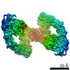
| Title | A two-site flexible clamp mechanism for RET-GDNF-GFRα1 assembly reveals both conformational adaptation and strict geometric spacing. |
|---|---|
| Journal, issue, pages | Structure, Vol. 29, Issue 7, Page 694-708.e7, Year 2021 |
| Publish date | Jul 1, 2021 |
 Authors Authors | Sarah E Adams / Andrew G Purkiss / Phillip P Knowles / Andrea Nans / David C Briggs / Annabel Borg / Christopher P Earl / Kerry M Goodman / Agata Nawrotek / Aaron J Borg / Pauline B McIntosh / Francesca M Houghton / Svend Kjær / Neil Q McDonald /  |
| PubMed Abstract | RET receptor tyrosine kinase plays vital developmental and neuroprotective roles in metazoans. GDNF family ligands (GFLs) when bound to cognate GFRα co-receptors recognize and activate RET ...RET receptor tyrosine kinase plays vital developmental and neuroprotective roles in metazoans. GDNF family ligands (GFLs) when bound to cognate GFRα co-receptors recognize and activate RET stimulating its cytoplasmic kinase function. The principles for RET ligand-co-receptor recognition are incompletely understood. Here, we report a crystal structure of the cadherin-like module (CLD1-4) from zebrafish RET revealing interdomain flexibility between CLD2 and CLD3. Comparison with a cryo-electron microscopy structure of a ligand-engaged zebrafish RET-GDNF-GFRα1a complex indicates conformational changes within a clade-specific CLD3 loop adjacent to the co-receptor. Our observations indicate that RET is a molecular clamp with a flexible calcium-dependent arm that adapts to different GFRα co-receptors, while its rigid arm recognizes a GFL dimer to align both membrane-proximal cysteine-rich domains. We also visualize linear arrays of RET-GDNF-GFRα1a suggesting that a conserved contact stabilizes higher-order species. Our study reveals that ligand-co-receptor recognition by RET involves both receptor plasticity and strict spacing of receptor dimers by GFL ligands. |
 External links External links | Structure / PubMed:33484636 / PubMed Central |
| Methods | EM (single particle) / X-ray diffraction |
| Resolution | 2.2 - 26.0 Å |
| Structure data |  EMDB-11777: EMDB-11822, PDB-7aml:  PDB-7ab8:  PDB-7amk: |
| Chemicals |  ChemComp-EDO:  ChemComp-PEG:  ChemComp-HOH:  ChemComp-CA:  ChemComp-NAG:  ChemComp-GOL:  ChemComp-PGE:  ChemComp-MES: |
| Source |
|
 Keywords Keywords |  SIGNALING PROTEIN / VERTEBRATE DEVELOPMENT / PART OF THE RET-GFL-GFRA COMPLEX / NEUROTROPHIC FACTOR / Ligand recognition / receptor tyrosine kinase / glycosylation SIGNALING PROTEIN / VERTEBRATE DEVELOPMENT / PART OF THE RET-GFL-GFRA COMPLEX / NEUROTROPHIC FACTOR / Ligand recognition / receptor tyrosine kinase / glycosylation |