ムービー
ムービー コントローラー
 構造ビューア
 万見文献について


+検索条件
-Structure paper
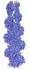
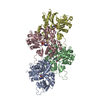
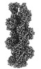
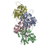
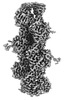
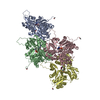
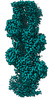
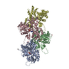
| タイトル | Structural insights into actin isoforms. |
|---|---|
| ジャーナル・号・ページ | Elife, Vol. 12, Year 2023 |
| 掲載日 | 2023年2月15日 |
 著者 著者 | Amandeep S Arora / Hsiang-Ling Huang / Ramanpreet Singh / Yoshie Narui / Andrejus Suchenko / Tomoyuki Hatano / Sarah M Heissler / Mohan K Balasubramanian / Krishna Chinthalapudi /   |
| PubMed 要旨 | Actin isoforms organize into distinct networks that are essential for the normal function of eukaryotic cells. Despite a high level of sequence and structure conservation, subtle differences in their ...Actin isoforms organize into distinct networks that are essential for the normal function of eukaryotic cells. Despite a high level of sequence and structure conservation, subtle differences in their design principles determine the interaction with myosin motors and actin-binding proteins. Therefore, identifying how the structure of actin isoforms relates to function is important for our understanding of normal cytoskeletal physiology. Here, we report the high-resolution structures of filamentous skeletal muscle α-actin (3.37 Å), cardiac muscle α-actin (3.07 Å), ß-actin (2.99 Å), and γ-actin (3.38 Å) in the Mg·ADP state with their native post-translational modifications. The structures revealed isoform-specific conformations of the N-terminus that shift closer to the filament surface upon myosin binding, thereby establishing isoform-specific interfaces. Collectively, the structures of single-isotype, post-translationally modified bare skeletal muscle α-actin, cardiac muscle α-actin, ß-actin, and γ-actin reveal general principles, similarities, and differences between isoforms. They complement the repertoire of known actin structures and allow for a comprehensive understanding of in vitro and in vivo functions of actin isoforms. |
 リンク リンク | Elife / PubMed:36790143 / PubMed Central |
| 手法 | EM (らせん対称) / EM (単粒子) |
| 解像度 | 2.99 - 3.38 Å |
| 構造データ | EMDB-27548, PDB-8dmx: EMDB-27549, PDB-8dmy: EMDB-27565, PDB-8dnf: EMDB-27572, PDB-8dnh: |
| 化合物 |  ChemComp-MG:  ChemComp-ADP: |
| 由来 |
|
 キーワード キーワード |  STRUCTURAL PROTEIN (タンパク質) / cytoskeleton (細胞骨格) STRUCTURAL PROTEIN (タンパク質) / cytoskeleton (細胞骨格) |