Movie
Movie Controller


+ Open data

- Basic information
| Entry |  | |||||||||
|---|---|---|---|---|---|---|---|---|---|---|
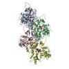
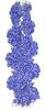
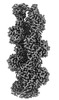
| Title | Cryo-EM structure of nonmuscle gamma-actin | |||||||||
 Map data Map data | Cryo-EM structure of nonmuscle gamma-actin | |||||||||
 Sample Sample |
| |||||||||
| Function / homology |  Function and homology information Function and homology informationbasal body patch /  tight junction assembly / regulation of transepithelial transport / structural constituent of postsynaptic actin cytoskeleton / protein localization to bicellular tight junction / morphogenesis of a polarized epithelium / profilin binding / Formation of annular gap junctions / Gap junction degradation / dense body ...basal body patch / tight junction assembly / regulation of transepithelial transport / structural constituent of postsynaptic actin cytoskeleton / protein localization to bicellular tight junction / morphogenesis of a polarized epithelium / profilin binding / Formation of annular gap junctions / Gap junction degradation / dense body / Cell-extracellular matrix interactions / regulation of stress fiber assembly / Adherens junctions interactions / Sensory processing of sound by outer hair cells of the cochlea / Interaction between L1 and Ankyrins / Sensory processing of sound by inner hair cells of the cochlea / sarcomere organization / NuA4 histone acetyltransferase complex / regulation of synaptic vesicle endocytosis / apical junction complex / regulation of focal adhesion assembly / maintenance of blood-brain barrier / positive regulation of wound healing / myofibril / Recycling pathway of L1 / filamentous actin / calyx of Held / EPH-ephrin mediated repulsion of cells / RHO GTPases Activate WASPs and WAVEs / RHO GTPases activate IQGAPs / RHOBTB2 GTPase cycle / phagocytic vesicle / EPHB-mediated forward signaling / axonogenesis / actin filament / cell motility / RHO GTPases Activate Formins / Translocation of SLC2A4 (GLUT4) to the plasma membrane / FCGR3A-mediated phagocytosis / Hydrolases; Acting on acid anhydrides; Acting on acid anhydrides to facilitate cellular and subcellular movement / Signaling by high-kinase activity BRAF mutants / Schaffer collateral - CA1 synapse / MAP2K and MAPK activation / structural constituent of cytoskeleton / Regulation of actin dynamics for phagocytic cup formation / platelet aggregation / VEGFA-VEGFR2 Pathway / cellular response to type II interferon / Signaling by RAF1 mutants / Signaling by moderate kinase activity BRAF mutants / Paradoxical activation of RAF signaling by kinase inactive BRAF / Signaling downstream of RAS mutants / cell-cell junction / Signaling by BRAF and RAF1 fusions / Clathrin-mediated endocytosis / angiogenesis / blood microparticle / cytoskeleton / hydrolase activity / positive regulation of cell migration / axon / focal adhesion / synapse / ubiquitin protein ligase binding / positive regulation of gene expression / protein kinase binding / extracellular space / extracellular exosome / ATP binding / membrane / identical protein binding / nucleus / plasma membrane / cytosol / cytoplasm tight junction assembly / regulation of transepithelial transport / structural constituent of postsynaptic actin cytoskeleton / protein localization to bicellular tight junction / morphogenesis of a polarized epithelium / profilin binding / Formation of annular gap junctions / Gap junction degradation / dense body ...basal body patch / tight junction assembly / regulation of transepithelial transport / structural constituent of postsynaptic actin cytoskeleton / protein localization to bicellular tight junction / morphogenesis of a polarized epithelium / profilin binding / Formation of annular gap junctions / Gap junction degradation / dense body / Cell-extracellular matrix interactions / regulation of stress fiber assembly / Adherens junctions interactions / Sensory processing of sound by outer hair cells of the cochlea / Interaction between L1 and Ankyrins / Sensory processing of sound by inner hair cells of the cochlea / sarcomere organization / NuA4 histone acetyltransferase complex / regulation of synaptic vesicle endocytosis / apical junction complex / regulation of focal adhesion assembly / maintenance of blood-brain barrier / positive regulation of wound healing / myofibril / Recycling pathway of L1 / filamentous actin / calyx of Held / EPH-ephrin mediated repulsion of cells / RHO GTPases Activate WASPs and WAVEs / RHO GTPases activate IQGAPs / RHOBTB2 GTPase cycle / phagocytic vesicle / EPHB-mediated forward signaling / axonogenesis / actin filament / cell motility / RHO GTPases Activate Formins / Translocation of SLC2A4 (GLUT4) to the plasma membrane / FCGR3A-mediated phagocytosis / Hydrolases; Acting on acid anhydrides; Acting on acid anhydrides to facilitate cellular and subcellular movement / Signaling by high-kinase activity BRAF mutants / Schaffer collateral - CA1 synapse / MAP2K and MAPK activation / structural constituent of cytoskeleton / Regulation of actin dynamics for phagocytic cup formation / platelet aggregation / VEGFA-VEGFR2 Pathway / cellular response to type II interferon / Signaling by RAF1 mutants / Signaling by moderate kinase activity BRAF mutants / Paradoxical activation of RAF signaling by kinase inactive BRAF / Signaling downstream of RAS mutants / cell-cell junction / Signaling by BRAF and RAF1 fusions / Clathrin-mediated endocytosis / angiogenesis / blood microparticle / cytoskeleton / hydrolase activity / positive regulation of cell migration / axon / focal adhesion / synapse / ubiquitin protein ligase binding / positive regulation of gene expression / protein kinase binding / extracellular space / extracellular exosome / ATP binding / membrane / identical protein binding / nucleus / plasma membrane / cytosol / cytoplasmSimilarity search - Function | |||||||||
| Biological species |  Homo sapiens (human) Homo sapiens (human) | |||||||||
| Method | single particle reconstruction / cryo EM / Resolution: 3.38 Å | |||||||||
 Authors Authors | Arora AS / Huang HL / Heissler SM / Chinthalapudi K | |||||||||
| Funding support |  United States, 1 items United States, 1 items
| |||||||||
 Citation Citation | Journal: Elife / Year: 2023 Title: Structural insights into actin isoforms. Authors: Amandeep S Arora / Hsiang-Ling Huang / Ramanpreet Singh / Yoshie Narui / Andrejus Suchenko / Tomoyuki Hatano / Sarah M Heissler / Mohan K Balasubramanian / Krishna Chinthalapudi /  Abstract: Actin isoforms organize into distinct networks that are essential for the normal function of eukaryotic cells. Despite a high level of sequence and structure conservation, subtle differences in their ...Actin isoforms organize into distinct networks that are essential for the normal function of eukaryotic cells. Despite a high level of sequence and structure conservation, subtle differences in their design principles determine the interaction with myosin motors and actin-binding proteins. Therefore, identifying how the structure of actin isoforms relates to function is important for our understanding of normal cytoskeletal physiology. Here, we report the high-resolution structures of filamentous skeletal muscle α-actin (3.37 Å), cardiac muscle α-actin (3.07 Å), ß-actin (2.99 Å), and γ-actin (3.38 Å) in the Mg·ADP state with their native post-translational modifications. The structures revealed isoform-specific conformations of the N-terminus that shift closer to the filament surface upon myosin binding, thereby establishing isoform-specific interfaces. Collectively, the structures of single-isotype, post-translationally modified bare skeletal muscle α-actin, cardiac muscle α-actin, ß-actin, and γ-actin reveal general principles, similarities, and differences between isoforms. They complement the repertoire of known actin structures and allow for a comprehensive understanding of in vitro and in vivo functions of actin isoforms. | |||||||||
| History |
|
- Structure visualization
| Supplemental images |
|---|

- Downloads & links
-EMDB archive
| Map data | emd_27565.map.gz | 59.7 MB | EMDB map data format | |
|---|---|---|---|---|
| Header (meta data) | emd-27565-v30.xmlemd-27565.xml | 17.1 KB 17.1 KB | Display Display | EMDB header |
| FSC (resolution estimation) | emd_27565_fsc.xml | 8.4 KB | Display | FSC data file |
| Images | 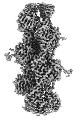 emd_27565.png emd_27565.png | 90.5 KB | ||
| Others | emd_27565_half_map_1.map.gzemd_27565_half_map_2.map.gz | 59.5 MB 59.5 MB | ||
| Archive directory |  http://ftp.pdbj.org/pub/emdb/structures/EMD-27565ftp://ftp.pdbj.org/pub/emdb/structures/EMD-27565 http://ftp.pdbj.org/pub/emdb/structures/EMD-27565ftp://ftp.pdbj.org/pub/emdb/structures/EMD-27565 | HTTPS FTP |
-Related structure data
| Related structure data | 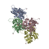 8dnfMC 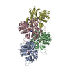 8dmxC 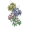 8dmyC 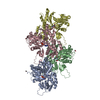 8dnhC M: atomic model generated by this map C: citing same article ( |
|---|---|
| Similar structure data |
-Links
| EMDB pages | EMDB (EBI/PDBe) / EMDataResource |
|---|---|
| Related items in Molecule of the Month |


-Map
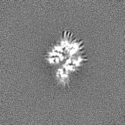
| File | Download / File: emd_27565.map.gz / Format: CCP4 / Size: 64 MB / Type: IMAGE STORED AS FLOATING POINT NUMBER (4 BYTES) | ||||||||||||||||||||
|---|---|---|---|---|---|---|---|---|---|---|---|---|---|---|---|---|---|---|---|---|---|
| Annotation | Cryo-EM structure of nonmuscle gamma-actin | ||||||||||||||||||||
| Voxel size | X=Y=Z: 0.891 Å | ||||||||||||||||||||
| Density |
| ||||||||||||||||||||
| Symmetry | Space group: 1 | ||||||||||||||||||||
| Details | EMDB XML:
|
-Supplemental data
-Half map: Cryo-EM structure of nonmuscle gamma-actin
| File | emd_27565_half_map_1.map | ||||||||||||
|---|---|---|---|---|---|---|---|---|---|---|---|---|---|
| Annotation | Cryo-EM structure of nonmuscle gamma-actin | ||||||||||||
| Projections & Slices |
| ||||||||||||
| Density Histograms |
 Z
Z Y
Y X
X


-Half map: Cryo-EM structure of nonmuscle gamma-actin
| File | emd_27565_half_map_2.map | ||||||||||||
|---|---|---|---|---|---|---|---|---|---|---|---|---|---|
| Annotation | Cryo-EM structure of nonmuscle gamma-actin | ||||||||||||
| Projections & Slices |
| ||||||||||||
| Density Histograms |


- Sample components
-Entire : actin
| Entire | Name: actin |
|---|---|
| Components |
|
-Supramolecule #1: actin
| Supramolecule | Name: actin / type: organelle_or_cellular_component / ID: 1 / Parent: 0 / Macromolecule list: #1 |
|---|---|
| Source (natural) | Organism: Homo sapiens (human) |
-Macromolecule #1: Actin, cytoplasmic 2, N-terminally processed
| Macromolecule | Name: Actin, cytoplasmic 2, N-terminally processed / type: protein_or_peptide / ID: 1 / Number of copies: 4 / Enantiomer: LEVO |
|---|---|
| Source (natural) | Organism: Homo sapiens (human) |
| Molecular weight | Theoretical: 41.746625 KDa |
| Recombinant expression | Organism:  Pichia (fungus) Pichia (fungus) |
| Sequence | String: (ACE)EEEIAALVI DNGSGMCKAG FAGDDAPRAV FPSIVGRPRH QGVMVGMGQK DSYVGDEAQS KRGILTLKYP IE (HIC)GIVTNW DDMEKIWHHT FYNELRVAPE EHPVLLTEAP LNPKANREKM TQIMFETFNT PAMYVAIQAV LSLYASGRT TGIVMDSGDG ...String: (ACE)EEEIAALVI DNGSGMCKAG FAGDDAPRAV FPSIVGRPRH QGVMVGMGQK DSYVGDEAQS KRGILTLKYP IE (HIC)GIVTNW DDMEKIWHHT FYNELRVAPE EHPVLLTEAP LNPKANREKM TQIMFETFNT PAMYVAIQAV LSLYASGRT TGIVMDSGDG VTHTVPIYEG YALPHAILRL DLAGRDLTDY LMKILTERGY SFTTTAEREI VRDIKEKLCY VALDFEQEMA TAASSSSLE KSYELPDGQV ITIGNERFRC PEALFQPSFL GMESCGIHET TFNSIMKCDV DIRKDLYANT VLSGGTTMYP G IADRMQKE ITALAPSTMK IKIIAPPERK YSVWIGGSIL ASLSTFQQMW ISKQEYDESG PSIVHRKCF |
-Macromolecule #2: MAGNESIUM ION
| Macromolecule | Name: MAGNESIUM ION / type: ligand / ID: 2 / Number of copies: 4 / Formula: MG |
|---|---|
| Molecular weight | Theoretical: 24.305 Da |
-Macromolecule #3: ADENOSINE-5'-DIPHOSPHATE
| Macromolecule | Name: ADENOSINE-5'-DIPHOSPHATE / type: ligand / ID: 3 / Number of copies: 4 / Formula: ADP |
|---|---|
| Molecular weight | Theoretical: 427.201 Da |
| Chemical component information |  ChemComp-ADP: |
-Experimental details
-Structure determination
| Method | cryo EM |
|---|---|
 Processing Processing | single particle reconstruction |
| Aggregation state | filament |
-Sample preparation
| Buffer | pH: 7 |
|---|---|
| Grid | Model: C-flat / Material: GOLD / Support film - Material: CARBON / Support film - topology: CONTINUOUS |
| Vitrification | Cryogen name: ETHANE / Chamber humidity: 95 % / Chamber temperature: 298 K / Instrument: LEICA EM GP |
- Electron microscopy
| Microscope | FEI TITAN KRIOS |
|---|---|
| Electron beam | Acceleration voltage: 300 kV / Electron source: FIELD EMISSION GUN |
| Electron optics | Illumination mode: OTHER / Imaging mode: BRIGHT FIELDBright-field microscopy / Nominal defocus max: 2.0 µm / Nominal defocus min: 0.3 µm / Nominal magnification: 81000 |
| Specialist optics | Spherical aberration corrector: Cs-corrected microscope / Energy filter - Name: GIF Bioquantum / Energy filter - Slit width: 20 eV |
| Sample stage | Specimen holder model: FEI TITAN KRIOS AUTOGRID HOLDER / Cooling holder cryogen: NITROGEN |
| Image recording | Film or detector model: GATAN K3 BIOQUANTUM (6k x 4k) / Number grids imaged: 1 / Number real images: 2952 / Average electron dose: 65.0 e/Å2 |
| Experimental equipment |  Model: Titan Krios / Image courtesy: FEI Company |
-Image processing
| Particle selection | Details: Single Particle |
|---|---|
| Startup model | Type of model: PDB ENTRY PDB model - PDB ID: |
| Initial angle assignment | Type: NOT APPLICABLE / Software - Name: cryoSPARC |
| Final angle assignment | Type: NOT APPLICABLE / Software - Name: cryoSPARC |
| Final reconstruction | Applied symmetry - Point group: C1 (asymmetric) / Resolution.type: BY AUTHOR / Resolution: 3.38 Å / Resolution method: FSC 0.143 CUT-OFF / Software - Name: cryoSPARC / Number images used: 1009372 |
| FSC plot (resolution estimation) |  |