 ムービー
ムービー コントローラー
 構造ビューア
 万見文献について


+検索条件
-Structure paper
| タイトル | Structural insights into the cause of human primary ciliary dyskinesia. |
|---|---|
| ジャーナル・号・ページ | Mol Biol Cell, Vol. 32, Issue 12, Page 1202-1209, Year 2021 |
| 掲載日 | 2021年6月1日 |
 著者 著者 | Yanhe Zhao / Justine Pinskey / Jianfeng Lin / Weining Yin / Patrick R Sears / Leigh A Daniels / Maimoona A Zariwala / Michael R Knowles / Lawrence E Ostrowski / Daniela Nicastro /  |
| PubMed 要旨 | Cilia and flagella are evolutionarily conserved eukaryotic organelles involved in cell motility and signaling. In humans, mutations in Radial Spoke Head Component 4A () can lead to primary ciliary ...Cilia and flagella are evolutionarily conserved eukaryotic organelles involved in cell motility and signaling. In humans, mutations in Radial Spoke Head Component 4A () can lead to primary ciliary dyskinesia (PCD), a life-shortening disease characterized by chronic respiratory tract infections, abnormal organ positioning, and infertility. Despite its importance for human health, the location of RSPH4A in human cilia has not been resolved, and the structural basis of PCD remains elusive. Here, we present the native three-dimensional structure of human respiratory cilia using samples collected noninvasively from a PCD patient. Using cryo-electron tomography (cryo-ET) and subtomogram averaging, we compared the structures of control and cilia, revealing primary defects in two of the three radial spokes (RSs) within the axonemal repeat and secondary (heterogeneous) defects in the central pair complex. Similar to cilia, the radial spoke heads of RS1 and RS2, but not RS3, were missing in cilia. However, cilia also exhibited defects within the arch domains adjacent to the RS1 and RS2 heads, which were not observed with RSPH1 loss. Our results provide insight into the underlying structural basis for PCD and highlight the benefits of applying cryo-ET directly to patient samples for molecular structure determination. |
 リンク リンク | Mol Biol Cell / PubMed:33852348 / PubMed Central |
| 手法 | EM (サブトモグラム平均) |
| 解像度 | 33.6 - 41.8 Å |
| 構造データ | 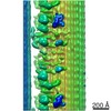 EMDB-22874: 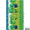 EMDB-22875: |
| 由来 |
|

 Homo sapiens (ヒト)