National Institutes of Health/National Institute of General Medical Sciences (NIH/NIGMS)
GM083122
United States
National Institutes of Health/National Institute of General Medical Sciences (NIH/NIGMS)
F32GM137470
United States
Cystic Fibrosis Foundation
R026-CR11
United States
National Institutes of Health/National Institute of General Medical Sciences (NIH/NIGMS)
DK065988
United States
National Institutes of Health/National Institute of General Medical Sciences (NIH/NIGMS)
HL117836
United States
National Institutes of Health/National Institute of General Medical Sciences (NIH/NIGMS)
HL096458
United States
National Institutes of Health/National Institute of General Medical Sciences (NIH/NIGMS)
HL071798
United States
Citation
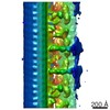
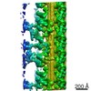
Journal: Mol Biol Cell / Year: 2021 Title: Structural insights into the cause of human primary ciliary dyskinesia. Authors: Yanhe Zhao / Justine Pinskey / Jianfeng Lin / Weining Yin / Patrick R Sears / Leigh A Daniels / Maimoona A Zariwala / Michael R Knowles / Lawrence E Ostrowski / Daniela Nicastro / Abstract: Cilia and flagella are evolutionarily conserved eukaryotic organelles involved in cell motility and signaling. In humans, mutations in Radial Spoke Head Component 4A () can lead to primary ciliary ...Cilia and flagella are evolutionarily conserved eukaryotic organelles involved in cell motility and signaling. In humans, mutations in Radial Spoke Head Component 4A () can lead to primary ciliary dyskinesia (PCD), a life-shortening disease characterized by chronic respiratory tract infections, abnormal organ positioning, and infertility. Despite its importance for human health, the location of RSPH4A in human cilia has not been resolved, and the structural basis of PCD remains elusive. Here, we present the native three-dimensional structure of human respiratory cilia using samples collected noninvasively from a PCD patient. Using cryo-electron tomography (cryo-ET) and subtomogram averaging, we compared the structures of control and cilia, revealing primary defects in two of the three radial spokes (RSs) within the axonemal repeat and secondary (heterogeneous) defects in the central pair complex. Similar to cilia, the radial spoke heads of RS1 and RS2, but not RS3, were missing in cilia. However, cilia also exhibited defects within the arch domains adjacent to the RS1 and RS2 heads, which were not observed with RSPH1 loss. Our results provide insight into the underlying structural basis for PCD and highlight the benefits of applying cryo-ET directly to patient samples for molecular structure determination.
History
Deposition
Oct 19, 2020
-
Header (metadata) release
May 5, 2021
-
Map release
May 5, 2021
-
Update
Jun 9, 2021
-
Current status
Jun 9, 2021
Processing site: RCSB / Status: Released
-
Structure visualization
Movie
Surface view with section colored by density value
In the structure databanks used in Yorodumi, some data are registered as the other names, "COVID-19 virus" and "2019-nCoV". Here are the details of the virus and the list of structure data.
Jan 31, 2019. EMDB accession codes are about to change! (news from PDBe EMDB page)
EMDB accession codes are about to change! (news from PDBe EMDB page)
The allocation of 4 digits for EMDB accession codes will soon come to an end. Whilst these codes will remain in use, new EMDB accession codes will include an additional digit and will expand incrementally as the available range of codes is exhausted. The current 4-digit format prefixed with “EMD-” (i.e. EMD-XXXX) will advance to a 5-digit format (i.e. EMD-XXXXX), and so on. It is currently estimated that the 4-digit codes will be depleted around Spring 2019, at which point the 5-digit format will come into force.
The EM Navigator/Yorodumi systems omit the EMD- prefix.
Related info.:Q: What is EMD? / ID/Accession-code notation in Yorodumi/EM Navigator
Yorodumi is a browser for structure data from EMDB, PDB, SASBDB, etc.
This page is also the successor to EM Navigator detail page, and also detail information page/front-end page for Omokage search.
The word "yorodu" (or yorozu) is an old Japanese word meaning "ten thousand". "mi" (miru) is to see.
Related info.:EMDB / PDB / SASBDB / Comparison of 3 databanks / Yorodumi Search / Aug 31, 2016. New EM Navigator & Yorodumi / Yorodumi Papers / Jmol/JSmol / Function and homology information / Changes in new EM Navigator and Yorodumi
 Movie
Movie Controller

 Yorodumi
 Open data
 Basic information
 Map data
 Sample
 Homo sapiens (human)
 Authors
 United States, 7 items
  Citation
 Structure visualization
 Movie viewer
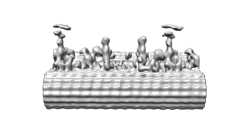
 Downloads & links
 emd_22874.png
 http://ftp.pdbj.org/pub/emdb/structures/EMD-22874
 Sample components
 Processing
 Electron microscopy
