 Movie
Movie Controller
 Structure viewers
 About EMN search


-Search query
-Search result
Showing 1 - 50 of 326 items for (author: gonen & t)

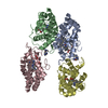
EMDB-45623: 
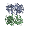
MicroED structure of the C11 cysteine protease clostripain
Method: electron crystallography / : Ruma YN, Bu G, Hattne J, Gonen T
PDB-9cip:
MicroED structure of the C11 cysteine protease clostripain
Method: electron crystallography / : Ruma YN, Bu G, Hattne J, Gonen T

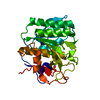
EMDB-43144:
MicroED structure of SARS-CoV-2 main protease (MPro/3CLPro) with missing cone eliminated by suspended drop
Method: electron crystallography / : Bu G, Gillman C, Danelius E, Hattne J, Nannenga BL, Gonen T
PDB-8vd7:
MicroED structure of SARS-CoV-2 main protease (MPro/3CLPro) with missing cone eliminated by suspended drop
Method: electron crystallography / : Bu G, Gillman C, Danelius E, Hattne J, Nannenga BL, Gonen T
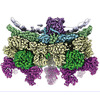
EMDB-43714:
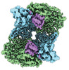
Cryo-EM structure of VP3-VP6 heterohexamer
Method: single particle / : Xia X, Sung PY, Martynowycz MW, Gonen T, Roy P, Zhou ZH

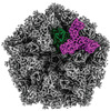
EMDB-43716:
Cryo-EM structure of BTV star-subcore
Method: single particle / : Xia X, Sung PY, Martynowycz MW, Gonen T, Roy P, Zhou ZH

EMDB-43719:
Cryo-EM structure of BTV pre-subcore
Method: single particle / : Xia X, Sung PY, Martynowycz MW, Gonen T, Roy P, Zhou ZH
EMDB-43722:
Cryo-EM structure of pre-subcore from in vitro assembled particles
Method: single particle / : Xia X, Sung PY, Martynowycz MW, Gonen T, Roy P, Zhou ZH
EMDB-43723:
Cryo-EM structure of BTV empty virion
Method: single particle / : Xia X, Sung PY, Martynowycz MW, Gonen T, Roy P, Zhou ZH
EMDB-43724:
Cryo-EM structure of BTV empty core
Method: single particle / : Xia X, Sung PY, Martynowycz MW, Gonen T, Roy P, Zhou ZH
EMDB-43725:
Cryo-EM structure of BTV empty pre-core
Method: single particle / : Xia X, Sung PY, Martynowycz MW, Gonen T, Roy P, Zhou ZH
EMDB-43726:
Cryo-EM structure of BTV subcore
Method: single particle / : Xia X, Sung PY, Martynowycz MW, Gonen T, Roy P, Zhou ZH
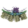
EMDB-43727:
Cryo-EM structure of BTV virion
Method: single particle / : Xia X, Sung PY, Martynowycz MW, Gonen T, Roy P, Zhou ZH
EMDB-43728:
Subtomogram averaging of BTV virion in host cells
Method: subtomogram averaging / : Xia X, Sung PY, Martynowycz MW, Gonen T, Roy P, Zhou ZH
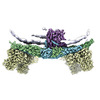
EMDB-43730:
Cryo-EM structure of BTV core
Method: single particle / : Xia X, Sung PY, Martynowycz MW, Gonen T, Roy P, Zhou ZH
EMDB-43731:
Cryo-EM structure of BTV pre-core
Method: single particle / : Xia X, Sung PY, Martynowycz MW, Gonen T, Roy P, Zhou ZH

PDB-8w12:
Cryo-EM structure of VP3-VP6 heterohexamer
Method: single particle / : Xia X, Sung PY, Martynowycz MW, Gonen T, Roy P, Zhou ZH

PDB-8w19:
Cryo-EM structure of BTV star-subcore
Method: single particle / : Xia X, Sung PY, Martynowycz MW, Gonen T, Roy P, Zhou ZH

PDB-8w1c:
Cryo-EM structure of BTV pre-subcore
Method: single particle / : Xia X, Sung PY, Martynowycz MW, Gonen T, Roy P, Zhou ZH

PDB-8w1i:
Cryo-EM structure of BTV subcore
Method: single particle / : Xia X, Sung PY, Martynowycz MW, Gonen T, Roy P, Zhou ZH

PDB-8w1o:
Cryo-EM structure of BTV virion
Method: single particle / : Xia X, Sung PY, Martynowycz MW, Gonen T, Roy P, Zhou ZH

PDB-8w1r:
Cryo-EM structure of BTV core
Method: single particle / : Xia X, Sung PY, Martynowycz MW, Gonen T, Roy P, Zhou ZH

PDB-8w1s:
Cryo-EM structure of BTV pre-core
Method: single particle / : Xia X, Sung PY, Martynowycz MW, Gonen T, Roy P, Zhou ZH

EMDB-28615:
MicroED structure of an Aeropyrum pernix protoglobin mutant
Method: electron crystallography / : Danelius E, Gonen T, Unge JT
PDB-8eum:
MicroED structure of an Aeropyrum pernix protoglobin mutant
Method: electron crystallography / : Danelius E, Gonen T, Unge JT

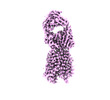
PDB-8skw:
MicroED structure of d(CGCGCG)2 Z-DNA
Method: electron crystallography / : Haymaker A, Bardin AA, Martynowycz MW, Nannenga BL

EMDB-28028:
I3-01 in an expanded conformation
Method: single particle / : McCarthy S, Gonen S

EMDB-28029:
I3-01 map refined in C1
Method: single particle / : McCarthy S, Gonen S

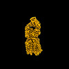
EMDB-40351:
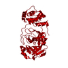
The MicroED structure of proteinase K crystallized by suspended drop crystallization
Method: electron crystallography / : Gillman C, Nicolas WJ, Martynowycz MW, Gonen T
PDB-8sdk:
The MicroED structure of proteinase K crystallized by suspended drop crystallization
Method: electron crystallography / : Gillman C, Nicolas WJ, Martynowycz MW, Gonen T

EMDB-29587:
MicroED structure of Proteinase K from lamellae milled from multiple plasma sources
Method: electron crystallography / : Martynowycz MW, Shiriaeva A, Clabbers MTB, Nicolas WJ, Weaver SJ, Hattne J, Gonen T

PDB-8fyo:
MicroED structure of Proteinase K from lamellae milled from multiple plasma sources
Method: electron crystallography / : Martynowycz MW, Shiriaeva A, Clabbers MTB, Nicolas WJ, Weaver SJ, Hattne J, Gonen T

EMDB-27148:
Zebrafish MFSD2A isoform B in inward open ligand bound conformation
Method: single particle / : Nguyen C, Lei HT, Lai LTF, Gallentino MJ, Mu X, Matthies D, Gonen T
EMDB-27149:
Zebrafish MFSD2A isoform B in inward open ligand-free conformation
Method: single particle / : Nguyen C, Lei HT, Lai LTF, Gallentino MJ, Mu X, Matthies D, Gonen T

EMDB-27150:
Zebrafish MFSD2A isoform B in inward open ligand 1A conformation
Method: single particle / : Nguyen C, Lei HT, Lai LTF, Gallentino MJ, Mu X, Matthies D, Gonen T

EMDB-27151:
Zebrafish MFSD2A isoform B in inward open ligand 1B conformation
Method: single particle / : Nguyen C, Lei HT, Lai LTF, Gallentino MJ, Mu X, Matthies D, Gonen T
EMDB-27152:
Zebrafish MFSD2A isoform B in inward open ligand 2B conformation
Method: single particle / : Nguyen C, Lei HT, Lai LTF, Gallentino MJ, Mu X, Matthies D, Gonen T
EMDB-27153:
Zebrafish MFSD2A isoform B in inward open ligand 3C conformation
Method: single particle / : Nguyen C, Lei HT, Lai LTF, Gallentino MJ, Mu X, Matthies D, Gonen T
PDB-8d2s:
Zebrafish MFSD2A isoform B in inward open ligand bound conformation
Method: single particle / : Nguyen C, Lei HT, Lai LTF, Gallentino MJ, Mu X, Matthies D, Gonen T
PDB-8d2t:
Zebrafish MFSD2A isoform B in inward open ligand-free conformation
Method: single particle / : Nguyen C, Lei HT, Lai LTF, Gallentino MJ, Mu X, Matthies D, Gonen T

PDB-8d2u:
Zebrafish MFSD2A isoform B in inward open ligand 1A conformation
Method: single particle / : Nguyen C, Lei HT, Lai LTF, Gallentino MJ, Mu X, Matthies D, Gonen T

PDB-8d2v:
Zebrafish MFSD2A isoform B in inward open ligand 1B conformation
Method: single particle / : Nguyen C, Lei HT, Lai LTF, Gallentino MJ, Mu X, Matthies D, Gonen T

PDB-8d2w:
Zebrafish MFSD2A isoform B in inward open ligand 2B conformation
Method: single particle / : Nguyen C, Lei HT, Lai LTF, Gallentino MJ, Mu X, Matthies D, Gonen T

PDB-8d2x:
Zebrafish MFSD2A isoform B in inward open ligand 3C conformation
Method: single particle / : Nguyen C, Lei HT, Lai LTF, Gallentino MJ, Mu X, Matthies D, Gonen T
EMDB-15244:
Tomogram of an Ebola VLP composed of GP, VP40, NP, VP24 and VP35 at pH 7.4 (Figure 1A-D)
Method: electron tomography / : Winter SL, Chlanda P
EMDB-15268:
Tomogram of an Ebola VLP composed of VP40 at pH 4.5 (Figure 1J)
Method: electron tomography / : Winter SL, Chlanda P

EMDB-15951:
Tomogram of an EBOV-infected Huh7 cell showing a late endosome with internalized EBOV particles
Method: electron tomography / : Winter SL, Chlanda P
EMDB-15956:
Tomogram of an extracellular EBOV particle adjacent to an EBOV-infected Huh7 cell
Method: electron tomography / : Winter SL, Chlanda P
EMDB-16128:
Tomogram of a late endosome of A549 cell infected with influenza A virus.
Method: electron tomography / : Klein S, Chlanda P
EMDB-16129:
Tomogram of a late endosome of A549 cell infected with influenza A virus (Figure 6C).
Method: electron tomography / : Klein S, Chlanda P
Pages:
 wwPDB to switch to version 3 of the EMDB data model
