 Movie
Movie Controller


+ Open data

- Basic information
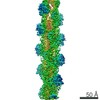
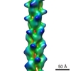
| Entry | Database: PDB / ID: 5kua | ||||||
|---|---|---|---|---|---|---|---|
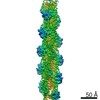
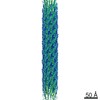
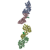
| Title | Cryo-EM reconstruction of Neisseria meningitidis Type IV pilus | ||||||
 Components Components | pilin | ||||||
 Keywords Keywords | PROTEIN FIBRIL / melted helix / Type IV pili | ||||||
| Function / homology |  Function and homology information Function and homology informationprotein secretion by the type II secretion system / type II protein secretion system complex / pilus / cell adhesion / membrane Similarity search - Function | ||||||
| Biological species |  Neisseria meningitidis (bacteria) Neisseria meningitidis (bacteria) | ||||||
| Method | ELECTRON MICROSCOPY / helical reconstruction / cryo EM / Resolution: 6 Å | ||||||
 Authors Authors | Kolappan, S. / Coureuil, M. / Yu, X. / Nassif, X. / Craig, L. / Egelman, E.H. | ||||||
 Citation Citation | Journal: Nat Commun / Year: 2016 Title: Structure of the Neisseria meningitidis Type IV pilus. Authors: Subramania Kolappan / Mathieu Coureuil / Xiong Yu / Xavier Nassif / Edward H Egelman / Lisa Craig /    Abstract: Neisseria meningitidis use Type IV pili (T4P) to adhere to endothelial cells and breach the blood brain barrier, causing cause fatal meningitis. T4P are multifunctional polymers of the major pilin ...Neisseria meningitidis use Type IV pili (T4P) to adhere to endothelial cells and breach the blood brain barrier, causing cause fatal meningitis. T4P are multifunctional polymers of the major pilin protein, which share a conserved hydrophobic N terminus that is a curved extended α-helix, α1, in X-ray crystal structures. Here we report a 1.44 Å crystal structure of the N. meningitidis major pilin PilE and a ∼6 Å cryo-electron microscopy reconstruction of the intact pilus, from which we built an atomic model for the filament. This structure reveals the molecular arrangement of the N-terminal α-helices in the filament core, including a melted central portion of α1 and a bridge of electron density consistent with a predicted salt bridge necessary for pilus assembly. This structure has important implications for understanding pilus biology. | ||||||
| History |
|
- Structure visualization
| Movie |
Movie viewer |
|---|---|
| Structure viewer | Molecule: MolmilJmol/JSmol |
- Downloads & links
-Download
| PDBx/mmCIF format | 5kua.cif.gz | 646.1 KB | Display | PDBx/mmCIF format |
|---|---|---|---|---|
| PDB format | pdb5kua.ent.gz | 538.5 KB | Display | PDB format |
| PDBx/mmJSON format | 5kua.json.gz | Tree view | PDBx/mmJSON format | |
| Others |  Other downloads Other downloads |
-Validation report
| Summary document | 5kua_validation.pdf.gz | 872.8 KB | Display | wwPDB validaton report |
|---|---|---|---|---|
| Full document | 5kua_full_validation.pdf.gz | 1.3 MB | Display | |
| Data in XML | 5kua_validation.xml.gz | 176.6 KB | Display | |
| Data in CIF | 5kua_validation.cif.gz | 234 KB | Display | |
| Arichive directory | https://data.pdbj.org/pub/pdb/validation_reports/ku/5kuaftp://data.pdbj.org/pub/pdb/validation_reports/ku/5kua | HTTPS FTP |
-Related structure data
| Related structure data |  8287MC  5jw8C M: map data used to model this data C: citing same article ( |
|---|---|
| Similar structure data |


-Links
 PDBj
PDBj
- Assembly
| Deposited unit | 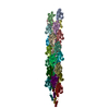
|
|---|---|
| 1 |
|
| Symmetry | Helical symmetry: (Circular symmetry: 1 / Dyad axis: no / N subunits divisor: 1 / Num. of operations: 26 / Rise per n subunits: 10.3 Å / Rotation per n subunits: 100.8 °) |
| Details | The biological unit is a helical fibril comprising an indeterminate number of identical subunits. The provided helical parameters should be applied to a single subunit to generate an extended fibril. |
-Components
| #1: Protein | Mass: 17067.182 Da / Num. of mol.: 26 / Source method: isolated from a natural source / Source: (natural) Neisseria meningitidis (bacteria) / References: UniProt: A0A1I9GEU1*PLUS |
|---|
-Experimental details
-Experiment
| Experiment | Method: ELECTRON MICROSCOPY |
|---|---|
| EM experiment | Aggregation state: FILAMENT / 3D reconstruction method: helical reconstruction |
- Sample preparation
| Component | Name: Type IV pilus / Type: ORGANELLE OR CELLULAR COMPONENT / Entity ID: all / Source: NATURAL |
|---|---|
| Source (natural) | Organism: Neisseria meningitidis (bacteria) |
| Buffer solution | pH: 10.5 |
| Specimen | Embedding applied: NO / Shadowing applied: NO / Staining applied: NO / Vitrification applied: YES |
| Vitrification | Instrument: FEI VITROBOT MARK IV / Cryogen name: ETHANE / Humidity: 90 % / Chamber temperature: 297 K / Details: Plunged into liquid ethane (FEI VITROBOT MARK IV) |
- Electron microscopy imaging
| Experimental equipment |  Model: Titan Krios / Image courtesy: FEI Company |
|---|---|
| Microscopy | Model: FEI TITAN KRIOS |
| Electron gun | Electron source:  FIELD EMISSION GUN / Accelerating voltage: 300 kV / Illumination mode: FLOOD BEAM FIELD EMISSION GUN / Accelerating voltage: 300 kV / Illumination mode: FLOOD BEAM |
| Electron lens | Mode: BRIGHT FIELD |
| Specimen holder | Cryogen: NITROGEN |
| Image recording | Electron dose: 40 e/Å2 / Film or detector model: FEI FALCON II (4k x 4k) |
- Processing
| Software | Name: PHENIX / Version: dev_2299: / Classification: refinement | ||||||||||||||||||||||||
|---|---|---|---|---|---|---|---|---|---|---|---|---|---|---|---|---|---|---|---|---|---|---|---|---|---|
| EM software | Name: SPIDER / Version: 13 / Category: 3D reconstruction | ||||||||||||||||||||||||
| CTF correction | Type: PHASE FLIPPING AND AMPLITUDE CORRECTION | ||||||||||||||||||||||||
| Helical symmerty | Angular rotation/subunit: 100.8 ° / Axial rise/subunit: 10.3 Å / Axial symmetry: C1 | ||||||||||||||||||||||||
| 3D reconstruction | Resolution: 6 Å / Resolution method: FSC 0.33 CUT-OFF / Num. of particles: 15586 / Algorithm: BACK PROJECTION / Symmetry type: HELICAL | ||||||||||||||||||||||||
| Atomic model building | Protocol: OTHER | ||||||||||||||||||||||||
| Refine LS restraints |
|
