 Movie
Movie Controller


+ Open data

- Basic information
| Entry | Database: PDB / ID: 2xkx | ||||||
|---|---|---|---|---|---|---|---|
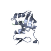
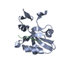
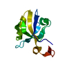
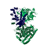
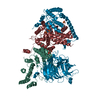
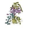
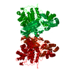
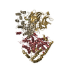
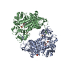
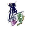
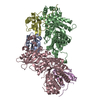
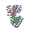
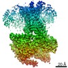
| Title | Single particle analysis of PSD-95 in negative stain | ||||||
 Components Components | DISKS LARGE HOMOLOG 4 | ||||||
 Keywords Keywords | STRUCTURAL PROTEIN / SCAFFOLD PROTEIN / MEMBRANE ASSOCIATED GUANYLATE KINASE | ||||||
| Function / homology |  Function and homology information Function and homology informationRHO GTPases activate CIT / positive regulation of AMPA glutamate receptor clustering / neuronal ion channel clustering / P2Y1 nucleotide receptor binding / Neurexins and neuroligins / beta-1 adrenergic receptor binding / neuroligin family protein binding / structural constituent of postsynaptic density / proximal dendrite / positive regulation of neuron projection arborization ...RHO GTPases activate CIT / positive regulation of AMPA glutamate receptor clustering / neuronal ion channel clustering / P2Y1 nucleotide receptor binding / Neurexins and neuroligins / beta-1 adrenergic receptor binding / neuroligin family protein binding / structural constituent of postsynaptic density / proximal dendrite / positive regulation of neuron projection arborization / synaptic vesicle maturation / regulation of grooming behavior / receptor localization to synapse / cellular response to potassium ion / protein localization to synapse / cerebellar mossy fiber / vocalization behavior / LGI-ADAM interactions / Trafficking of AMPA receptors / neuron spine / dendritic branch / Activation of Ca-permeable Kainate Receptor / AMPA glutamate receptor clustering / frizzled binding / dendritic spine morphogenesis / juxtaparanode region of axon / establishment or maintenance of epithelial cell apical/basal polarity / negative regulation of receptor internalization / dendritic spine organization / postsynaptic neurotransmitter receptor diffusion trapping / neuron projection terminus / acetylcholine receptor binding / positive regulation of synapse assembly / regulation of NMDA receptor activity / Synaptic adhesion-like molecules / positive regulation of dendrite morphogenesis / RAF/MAP kinase cascade / beta-2 adrenergic receptor binding / neurotransmitter receptor localization to postsynaptic specialization membrane / cortical cytoskeleton / locomotory exploration behavior / regulation of neuronal synaptic plasticity / kinesin binding / extrinsic component of cytoplasmic side of plasma membrane / positive regulation of excitatory postsynaptic potential / AMPA glutamate receptor complex / social behavior / neuromuscular process controlling balance / positive regulation of protein tyrosine kinase activity / excitatory synapse / D1 dopamine receptor binding / Unblocking of NMDA receptors, glutamate binding and activation / positive regulation of synaptic transmission / glutamate receptor binding / ionotropic glutamate receptor binding / dendrite cytoplasm / synaptic membrane / cell periphery / PDZ domain binding / kinase binding / cell-cell adhesion / postsynaptic density membrane / regulation of long-term neuronal synaptic plasticity / establishment of protein localization / neuromuscular junction / cerebral cortex development / cell junction / cell-cell junction / synaptic vesicle / positive regulation of cytosolic calcium ion concentration / basolateral plasma membrane / protein phosphatase binding / scaffold protein binding / protein-containing complex assembly / chemical synaptic transmission / postsynaptic membrane / postsynapse / dendritic spine / postsynaptic density / neuron projection / signaling receptor binding / glutamatergic synapse / synapse / dendrite / protein-containing complex binding / protein kinase binding / endoplasmic reticulum / membrane / plasma membrane / cytoplasm / cytosol Similarity search - Function | ||||||
| Biological species |  | ||||||
| Method | ELECTRON MICROSCOPY /  SOLUTION SCATTERING / single particle reconstruction / negative staining / Resolution: 22.9 Å SOLUTION SCATTERING / single particle reconstruction / negative staining / Resolution: 22.9 Å | ||||||
| Model type details | CA ATOMS ONLY, CHAIN A, B | ||||||
 Authors Authors | Fomina, S. / Howard, T.D. / Sleator, O.K. / Golovanova, M. / O'Ryan, L. / Leyland, M.L. / Grossmann, J.G. / Collins, R.F. / Prince, S.M. | ||||||
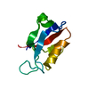
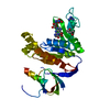
 Citation Citation | Journal: Biochim Biophys Acta / Year: 2011 Title: Self-directed assembly and clustering of the cytoplasmic domains of inwardly rectifying Kir2.1 potassium channels on association with PSD-95. Authors: Svetlana Fomina / Tina D Howard / Olivia K Sleator / Marina Golovanova / Liam O'Ryan / Mark L Leyland / J Günter Grossmann / Richard F Collins / Stephen M Prince /  Abstract: The interaction of the extra-membranous domain of tetrameric inwardly rectifying Kir2.1 ion channels (Kir2.1NC(4)) with the membrane associated guanylate kinase protein PSD-95 has been studied using ...The interaction of the extra-membranous domain of tetrameric inwardly rectifying Kir2.1 ion channels (Kir2.1NC(4)) with the membrane associated guanylate kinase protein PSD-95 has been studied using Transmission Electron Microscopy in negative stain. Three types of complexes were observed in electron micrographs corresponding to a 1:1 complex, a large self-enclosed tetrad complex and extended chains of linked channel domains. Using models derived from small angle X-ray scattering experiments in which high resolution structures from X-ray crystallographic and Nuclear Magnetic Resonance studies are positioned, the envelopes from single particle analysis can be resolved as a Kir2.1NC(4):PSD-95 complex and a tetrad of this unit (Kir2.1NC(4):PSD-95)(4). The tetrad complex shows the close association of the Kir2.1 cytoplasmic domains and the influence of PSD-95 mediated self-assembly on the clustering of these channels. #1: Journal: J Mol Biol / Year: 2003Title: Supramodular structure and synergistic target binding of the N-terminal tandem PDZ domains of PSD-95. Authors: Jia-Fu Long / Hidehito Tochio / Ping Wang / Jing-Song Fan / Carlo Sala / Martin Niethammer / Morgan Sheng / Mingjie Zhang /  Abstract: PDZ domain proteins play critical roles in binding, clustering and subcellular targeting of membrane receptors and ion channels. PDZ domains in multi-PDZ proteins often are arranged in groups with ...PDZ domain proteins play critical roles in binding, clustering and subcellular targeting of membrane receptors and ion channels. PDZ domains in multi-PDZ proteins often are arranged in groups with highly conserved spacing and intervening sequences; however, the functional significance of such tandem arrangements of PDZs is unclear. We have solved the three-dimensional structure of the first two PDZ domains of postsynaptic density protein-95 (PSD-95 PDZ1 and PDZ2), which are closely linked to each other in the PSD-95 family of scaffold proteins. The two PDZs have limited freedom of rotation and their C-terminal peptide-binding grooves are aligned with each other with an orientation preference for binding to pairs of C termini extending in the same direction. Increasing the spacing between PDZ1 and PDZ2 resulted in decreased binding between PDZ12 and its dimeric targets. The same mutation impaired the functional ability of PSD-95 to cluster Kv1.4 potassium channels in heterologous cells. The data presented provide a molecular basis for preferential binding of PSD-95 to multimeric membrane proteins with appropriate C-terminal sequences. #2: Journal: J Mol Biol / Year: 2000Title: Solution structure and backbone dynamics of the second PDZ domain of postsynaptic density-95. Authors: H Tochio / F Hung / M Li / D S Bredt / M Zhang / Abstract: The second PDZ domain of postsynaptic density-95 (PSD-95 PDZ2) plays a critical role in coupling N-methyl-D-aspartate receptors to neuronal nitric oxide synthase (nNOS). In this work, the solution ...The second PDZ domain of postsynaptic density-95 (PSD-95 PDZ2) plays a critical role in coupling N-methyl-D-aspartate receptors to neuronal nitric oxide synthase (nNOS). In this work, the solution structure of PSD-95 PDZ2 was determined to high resolution by NMR spectroscopy. The structure of PSD-95 PDZ2 was compared in detail with that of alpha1-syntrophin PDZ domain, as the PDZ domains share similar target interaction properties. The interaction of the PSD-95 PDZ2 with a carboxyl-terminal peptide derived from a cytoplasmic protein CAPON was studied by NMR titration experiments. Complex formation between PSD-95 PDZ2 and the nNOS PDZ was modelled on the basis of the crystal structure of the alpha1-syntrophin PDZ/nNOS PDZ dimer. We found that the prolonged loop connecting the betaB and betaC strands of PSD-95 PDZ2 is likely to play a role in both the binding of the carboxyl-terminal peptide and the nNOS beta-finger. Finally, the backbone dynamics of the PSD-95 PDZ2 in the absence of bound peptide were studied using a model-free approach. The "GLGF"-loop and the loop connecting alphaB and betaF of the protein display some degree of flexibility in solution. The rest of the protein is rigid and lacks detectable slow time-scale (microseconds to milliseconds) motions. In particular, the loop connecting betaB and betaC loop adopts a well-defined, rigid structure in solution. It appears that the loop adopts a pre-aligned conformation for the PDZ domain to interact with its targets. #3: Journal: To be PublishedTitle: Structure of the Third Pdz Domain of Psd-95 Protein Complexed with Kketwv Peptide Ligand Authors: Saro, D. / Wawrzak, Z. / Martin, P. / Vickrey, J. / Paredes, A. / Kovari, L. / Spaller, M. #4: Journal: Mol Cell / Year: 2001Title: Structure of the SH3-guanylate kinase module from PSD-95 suggests a mechanism for regulated assembly of MAGUK scaffolding proteins. Authors: A W McGee / S R Dakoji / O Olsen / D S Bredt / W A Lim / K E Prehoda /  Abstract: Membrane-associated guanylate kinases (MAGUKs), such as PSD-95, are modular scaffolds that organize signaling complexes at synapses and other cell junctions. MAGUKs contain PDZ domains, which recruit ...Membrane-associated guanylate kinases (MAGUKs), such as PSD-95, are modular scaffolds that organize signaling complexes at synapses and other cell junctions. MAGUKs contain PDZ domains, which recruit signaling proteins, as well as a Src homology 3 (SH3) and a guanylate kinase-like (GK) domain, implicated in scaffold oligomerization. The crystal structure of the SH3-GK module from PSD-95 reveals that these domains form an integrated unit: the SH3 fold comprises noncontiguous sequence elements divided by a hinge region and the GK domain. These elements compose two subdomains that can assemble in either an intra- or intermolecular fashion to complete the SH3 fold. We propose a model for MAGUK oligomerization in which complementary SH3 subdomains associate by 3D domain swapping. This model provides a possible mechanism for ligand regulation of oligomerization. | ||||||
| History |
|
- Structure visualization
| Movie |
Movie viewer |
|---|---|
| Structure viewer | Molecule: MolmilJmol/JSmol |
- Downloads & links
-Download
| PDBx/mmCIF format | 2xkx.cif.gz | 56.5 KB | Display | PDBx/mmCIF format |
|---|---|---|---|---|
| PDB format | pdb2xkx.ent.gz | 32.3 KB | Display | PDB format |
| PDBx/mmJSON format | 2xkx.json.gz | Tree view | PDBx/mmJSON format | |
| Others |  Other downloads Other downloads |
-Validation report
| Summary document | 2xkx_validation.pdf.gz | 739.9 KB | Display | wwPDB validaton report |
|---|---|---|---|---|
| Full document | 2xkx_full_validation.pdf.gz | 757.3 KB | Display | |
| Data in XML | 2xkx_validation.xml.gz | 25.5 KB | Display | |
| Data in CIF | 2xkx_validation.cif.gz | 35.8 KB | Display | |
| Arichive directory | https://data.pdbj.org/pub/pdb/validation_reports/xk/2xkxftp://data.pdbj.org/pub/pdb/validation_reports/xk/2xkx | HTTPS FTP |
-Related structure data
| Related structure data | 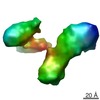 1761MC 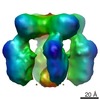 1764C 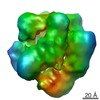 1765C 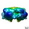 1766C 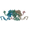 2xkyC C: citing same article ( M: map data used to model this data |
|---|---|
| Similar structure data |


-Links
 PDBj
PDBj


- Assembly
| Deposited unit | 
|
|---|---|
| 1 |
|
-Components
| #1: Protein | Mass: 80203.047 Da / Num. of mol.: 2 / Mutation: YES Source method: isolated from a genetically manipulated source Source: (gene. exp.)  Compound details | ENGINEERED RESIDUE IN CHAIN A, THR 9 TO ALA ENGINEERED RESIDUE IN CHAIN A, GLU 23 TO LYS ENGINEERED ...ENGINEERED | |
|---|
-Experimental details
-Experiment
| Experiment |
| |||
|---|---|---|---|---|
| EM experiment | Aggregation state: PARTICLE / 3D reconstruction method: single particle reconstruction |
- Sample preparation
| Component | Name: RAT PSD-95 / Type: COMPLEX |
|---|---|
| Buffer solution | Name: 20MM TRIS/HCL, 5MM DTT, 1MM EDTA, / pH: 7.5 / Details: 20MM TRIS/HCL, 5MM DTT, 1MM EDTA, |
| Specimen | Conc.: 1 mg/ml / Embedding applied: NO / Shadowing applied: NO / Staining applied: YES / Vitrification applied: NO |
| EM staining | Type: NEGATIVE / Material: Uranyl Acetate |
| Specimen support | Details: CARBON |
-Data collection
| Microscopy | Model: FEI TECNAI 10 / Details: LOW DOSE | |||||||||||||||||||||||||||||||||||||||
|---|---|---|---|---|---|---|---|---|---|---|---|---|---|---|---|---|---|---|---|---|---|---|---|---|---|---|---|---|---|---|---|---|---|---|---|---|---|---|---|---|
| Electron gun | Electron source: TUNGSTEN HAIRPIN / Accelerating voltage: 100 kV / Illumination mode: FLOOD BEAM | |||||||||||||||||||||||||||||||||||||||
| Electron lens | Mode: BRIGHT FIELD / Nominal magnification: 43000 X / Nominal defocus max: 2250 nm / Nominal defocus min: 900 nm / Cs: 3.6 mm | |||||||||||||||||||||||||||||||||||||||
| Specimen holder | Tilt angle max: 0.1 ° / Tilt angle min: 0 ° | |||||||||||||||||||||||||||||||||||||||
| Image recording | Film or detector model: GENERIC FILM | |||||||||||||||||||||||||||||||||||||||
| Image scans | Num. digital images: 2 | |||||||||||||||||||||||||||||||||||||||
| Radiation wavelength | Relative weight: 1 | |||||||||||||||||||||||||||||||||||||||
| Soln scatter |
|
- Processing
| EM software |
| |||||||||||||||||||||||||||||||||||
|---|---|---|---|---|---|---|---|---|---|---|---|---|---|---|---|---|---|---|---|---|---|---|---|---|---|---|---|---|---|---|---|---|---|---|---|---|
| CTF correction | Details: PARAMETERS DETERMINED USING SCATTERING CURVE | |||||||||||||||||||||||||||||||||||
| Symmetry | Point symmetry: C1 (asymmetric) | |||||||||||||||||||||||||||||||||||
| 3D reconstruction | Resolution: 22.9 Å / Num. of particles: 7854 / Nominal pixel size: 3.667 Å / Actual pixel size: 3.667 Å Details: THE COORDINATES DEPOSITED ARE FROM A COMBINED SAXS/EM STUDY. THE DOMAINS IN THE PROTEINS (HIGH RESOLUTION STRUCTURES FROM THE PDB) ARE POSITIONED RELATIVE TO ONE ANOTHER USING A SAXS CURVE, ...Details: THE COORDINATES DEPOSITED ARE FROM A COMBINED SAXS/EM STUDY. THE DOMAINS IN THE PROTEINS (HIGH RESOLUTION STRUCTURES FROM THE PDB) ARE POSITIONED RELATIVE TO ONE ANOTHER USING A SAXS CURVE, THIS COMPOSITE STRUCTURE IS THEN FITTED INTO AN EM MAP. DOMAINS PROVIDED BY X-RAY, NMR POSITIONED BY BUNCH ( PETOUKHOV, M. V. & SVERGUN, D. I. (2005). GLOBAL RIGID BODY MODELING OF MACROMOLECULAR COMPLEXES AGAINST SMALL- ANGLE SCATTERING DATA. BIOPHYS J 89, 1237-50.) REFINEMENT SUBMISSION BASED ON EXPERIMENTAL DATA FROM EMDB EMD-1761. (DEPOSITION ID: 7384). Symmetry type: POINT | |||||||||||||||||||||||||||||||||||
| Atomic model building | Protocol: RIGID BODY FIT / Space: REAL Details: METHOD--A MAP WAS GENERATED FROM THE SAXS MODEL COORDINATES AT A RESOLUTION MATCHING THE EXPERIMENTAL MAP. T HIS CALCULATED MAP WAS FITTED INTO THE EXPERIMENTAL MAP BY MAXIMIZING THE CROSS- ...Details: METHOD--A MAP WAS GENERATED FROM THE SAXS MODEL COORDINATES AT A RESOLUTION MATCHING THE EXPERIMENTAL MAP. T HIS CALCULATED MAP WAS FITTED INTO THE EXPERIMENTAL MAP BY MAXIMIZING THE CROSS-CORRELATION WITH THE EXPERIMENTAL MAP. THE COORDINATES WERE THEN REPLACED IN THE CALCULATED MAP TO GENERATE THE FINAL ENTRY. REFINEMENT PROTOCOL--DOCKED USING CHIMERA | |||||||||||||||||||||||||||||||||||
| Atomic model building |
| |||||||||||||||||||||||||||||||||||
| Refinement | Highest resolution: 22.9 Å | |||||||||||||||||||||||||||||||||||
| Refinement step | Cycle: LAST / Highest resolution: 22.9 Å
| |||||||||||||||||||||||||||||||||||
| Soln scatter model | Method: RIGID BODY MODELLING Conformer selection criteria: TWO MAJOR CONFORMERS WERE PRESENT IN THE POOL OF REFINED SAXS MODELS CHAIN A REPRESENTS THE BEST AGREEMENT WITH THE EXPERIMENTAL SAXS DATA (CHI=3.3). CHAIN B IS ...Conformer selection criteria: TWO MAJOR CONFORMERS WERE PRESENT IN THE POOL OF REFINED SAXS MODELS CHAIN A REPRESENTS THE BEST AGREEMENT WITH THE EXPERIMENTAL SAXS DATA (CHI=3.3). CHAIN B IS REPRESENTATIVE OF THE SECOND CONFORMATION (CHI=3.8). Details: NUMBER OF TIME FRAMES USED 24 (60S, 4.25M CAMERA), 40(60S, 1M CAMERA). PROTEIN CONCENTRATION 1 MG/ML (4.25M CAMERA) 8 MG/ML (1M CAMERA) Entry fitting list: PROGRAM PRE-BUNCH 1IU0/1QLC/1TP5/1KJW + SEQUENCE DATA Num. of conformers calculated: 20 / Num. of conformers submitted: 2 / Software author list: PETOUKHOV, M. V. & SVERGUN, D. I. / Software list: SASREF7/BUNCH8 |
